SynthePURE™ is a Whey Protein Isolate fashioned through a process performed at a much lower temperature than is typical for whey protein manufacture. During this process the protein powder is thoroughly filtered resulting in a high purity end product. The lower temperature has the effect of preserving the fragile biologically active peptides, lactoferrin, and immunoglobulins found in whey. The result is a whey protein powder devoid of denaturation with no loss of biological activity.
Protein is the essential centerpiece around which all other factors connect to promote anabolism. This article will focus on protein metabolism and the role the various hormones play in complementing each other in inducing anabolism. SynthePURE™ is the substrate upon which all of the factors work in effecting net muscle protein synthesis. We will examine in a highly readable (non-technical) manner the significance of:
- Insulin
- Growth Hormone
- Amino Acid Pool
- Exercise
- Blood Flow
- IGF-1
- IGF-1 bound to IGFBP-3
- Androgens
- Thyroid Hormones
None of these factors are capable of bringing about muscle tissue accrual by themselves. Instead these factors complement each other and do so by facilitating the reduction of the breakdown or promotion of the synthesis of muscle protein. Some factors are only capable of positively effecting retainment or uptake of a single crucial amino acid while others have multifaceted roles to play. Most of these factors are not anabolic by themselves. Rather they contribute specific necessities to a central pool that together maximize anabolism. All of these factors modulate amino acids which come from protein.
The Centerpiece – Protein
Quality
Infant formulas rely on bovine whey protein to mimic the nutritional content of human milk. Of critical importance in substituting for human milk is the inclusion of the highly nutritious alpha-Lactalbumin protein found in whey. Both bovine and human alpha-Lactalbumin contain very high amounts of the essential amino acids (tryptophan, phenylalanine + tyrosine, leucine, isoleucine, threonine, methionine + cysteine, lysine and valine). Of primary importance are the significant quantities of lysine, cysteine and tryptophan contained in alpha-Lactalbumin1. Unfortunately the processing through which protein undergoes in creating a humanly consumable product harms some of these amino acids. Tryptophan is a particularly unstable amino acid, and the heating of proteins at excessive temperatures will cause major reductions in tryptophan bioavailability 2. In addition the reaction between nitrogenous side chains of the amino acids and reducing sugars usually brings about a deterioration of the nutritional quality of the protein and lysine in particular is often lost. Lysine, tryptophan and methionine residues also react with oxidizing lipids and cause losses in the availability of lysine, tryptophan and sulfur-containing amino acids 3.
The important point is simply if loss of protein bioavailability is unacceptable for infant nutrition it should also be unacceptable to those seeking body transformation.
In the literature there has been considerable interest in the proposition that proteins of different biological quality and digestibility might be more or less efficient at supplying amino acids to muscle after exercise. Recent studies 4,5 seem to demonstrate that whey proteins are superior to casein and soy in supplying amino acids for net muscle protein accretion.
Quality protein is the centerpiece around which tissue accrual evolves.
Why is protein the centerpiece?
“The metabolic basis for skeletal muscle growth lies in the relationship of muscle protein synthesis to muscle protein breakdown. Muscle hypertrophy occurs only from net protein synthesis; that is, when muscle protein synthesis exceeds breakdown….resistance exercise, has a profound effect on muscle protein metabolism, often resulting in muscle growth. Acutely, resistance exercise may result in improved muscle protein balance, but, in the absence of food intake, the balance remains negative (i.e., catabolic). …Amino acid availability is critical to the control of muscle protein metabolism. Thus, a meal or a supplement containing protein or amino acids will influence muscle protein. …[and if] ingested after exercise should result in muscle anabolism.” – Kevin Tipton 6
Normally meals induce a transient increase in muscle protein synthesis (MPS) followed by muscle protein breakdown (MPB). In this manner tissue is maintained but there is no net protein synthesis thus no anabolism.

The authors of the Tipton study from which the above quote derives established therein that ingestion of either whey protein or casein protein after exercise led to “increases in muscle protein net balance, resulting in net muscle protein synthesis despite different patterns of blood amino acid responses” 6.
The general consensus from research in this area is that exercise induces protein degradation as well as protein synthesis. Ingestion of protein during this time period strongly tips the balance of degradation versus synthesis to that of overall protein synthesis.

There really is very little to be gained by spending a lot of time discussing the details of numerous studies which examine such things as:
- Digestion Rate: Whey versus Casein and EAA (essential amino acids); and
- Timing: pre-workout versus during and post-workout
Making Whey digest slowly
Most of the variability between the various protein sources lies in the digestion rate. Whey is a fast digesting protein and can be made to behave like the even speedier essential amino acids (EAA) by simply ingesting whey a little earlier. This is an effective pre-workout approach. Like-wise if a slower digestion rate is desired co-ingestion of viscous gels and soluble dietary fibers such as psyllium, pectin, guar gum and ispaghula will slow amino acid release either by increasing the time needed for intestinal absorption or slowing the rate of gastric emptying 7-11. In this manner whey can be made to behave as the slower releasing soy and casein proteins.
So while whey protein can be made to behave more like casein, casein can not be made to behave like whey.
Just ingest it
Exercise and whey protein are anabolic no matter when ingested. The effect whey protein has on anabolism can be explained through the microscope. A more comfortable approach though is to simply back away from all of the details many of which have yet to be elucidated and focus on a natural discussion of those familiar factors that ultimately influence protein metabolism at the microscopic level. That is the approach we will follow. But since we have the microscope in front of us lets take a quick look.
The regulation of skeletal muscle protein turnover is complex. It involves the interactions of gene transcription (i.e. obtaining the assembly instructions) and the subsequent translational control of protein synthesis (i.e. the assembly of the protein from its constituent parts, amino acids). The primary translation pathway leading to protein synthesis is mTOR (mammalian target of rapamycin). In order to build proteins, translation needs to occur and this is initiated by many factors and signaling molecules which feed into this complex regulator called mTOR.
mTOR is a key regulator of translational control. Nutrient, hormonal, and contractile stimuli primarily converge at this protein making mTOR an important modulator of protein synthesis. So when we back away from the microscope and discuss hormones such as insulin, IGF-1 and growth hormone and factors such as exercise, blood flow and protein ingestion what we should be vaguely aware of is that these hormones and factors are in part converging on mTOR to activate those positive regulators of mTOR and protein synthesis or deactivate those negative regulators (or inhibitors) of mTOR and subsequent protein synthesis.
There is no need to examine the specifics and so the following image is meant to convey a very general understanding of the complexity inside the cell.

A recent study in humans using muscle biopsies, tissue processing, western immunoblot analysis and maybe a microscope as well concluded that resistance exercise rapidly increases mTOR signaling, and whey protein increases and prolongs the mTOR signaling response to exercise and training 12.
Simply stated whey protein following exercise increases net protein synthesis which leads to anabolism. If you have a microscope you can add “through mTOR”.
References:
1 – Heine, Willi E., The Importance of alpha-Lactalbumin in Infant Nutrition, J. Nutr. 121: 277-283, 1991
2 – Cug, J. L. & Friedman, M. (1989), Effect of heat on tryptophan in food: chemistry, toxicology, and nutritional consequences Absorption and Utilization of Amino Acids, (Friedman, M., ed.), vol. 3, pp. 103-115, CRC Press, Boca Raton, FL
3 – Nielsen, H. K., Finot, P. A & Hurrell, R. F. (1985), Reactions of proteins with oxidizing lipids: 2. Influence on protein quality and on the bioavailability of lysine, methionine, cyst(e)ine and tryptophan as measured in rat assays, Br. J. Nutr. 53: 75-86
4 – Hartman JW, Tang JE, Wilkinson SB, Tarnopolsky MA, Lawrence RL, Fullerton AV, Phillips SM, Consumption of fat-free fluid milk after resistance exercise promotes greater lean mass accretion than does consumption of soy or carbohydrate in young, novice, male weightlifters, Am J Clin Nutr 86: 373–381, 2007
5 – Wilkinson SB, Tarnopolsky MA, Macdonald MJ, MacDonald JR, Armstrong D, Phillips SM, Consumption of fluid skim milk promotes greater muscle protein accretion after resistance exercise than does consumption of an isonitrogenous and isoenergetic soy-protein beverage, Am J Clin Nutr 85: 1031–1040, 2007
6 – Tipton, Kevin, et al., Ingestion of Casein and Whey Proteins Result in Muscle Anabolism after Resistance Exercise, Med Sci Sports Exerc. 2004 Dec;36(12):2073-81
7 – Rigaud, D., Effect of psyllium on gastric emptying, hunger feeling and food intake in normal volunteers: a double blind study, European Journal of Clinical Nutrition (1998) 52, 239-245
8 – Holt S, Heading RC, Cater DC, Prescott LF & Tothill P (1979): Effect of gel fibre on gastric emptying and absorption of glucose and paracetamol, Lancet 1, 636-639
9 – Blackburn NA, Redfern JS, Jarjis HA, Holgate AM, Hanning I & Scarpello JH (1984): The mechanism of action of guar gum in improving glucose tolerance in man, Clin. Sci. 66, 329-36
10 – Ralphs DNL & Lawaetz NJG (1978): Effect of a dietary fibre on gastric emptying in dumpers, Gut 19, A 986-987
11 – Schwartz SE, Levine RA, Singh A, Scheidecker JR & Track NS (1982): Sustained pectin ingestion delays gastric emptying, Gastroentero. 83, 812-817
12 – Hulmi, J. J., Resistance exercise with whey protein ingestion affects mTOR signaling pathway and myostatin in men, Appl Physiol 106: 1720–1729, 2009
Protein Metabolism – How various hormones create anabolism.
A complete protein such as SynthePURE™ provides the raw materials (amino acids) for protein synthesis. We understand that anabolism only occurs when protein synthesis exceeds protein degradation/breakdown. Exercise creates an environment where anabolism is possible if protein is supplied. In that environment protein synthesis will exceed protein breakdown resulting in net tissue accrual.
However ultimate body transformation requires maximum anabolism. This is primarily achieved through the complementary interplay of those hormones that affect the components of protein metabolism. By understanding precisely how each hormone or factor affects the components responsible for the outcome of protein metabolism one can better achieve an anabolic response.
The primary components responsible for determining the outcome of protein metabolism are:
- Whole Body Protein Synthesis – synthesis throughout organs, non skeletal muscle & skeletal muscle
- Muscle Protein Synthesis – synthesis in skeletal muscle only
- Whole Body Protein Breakdown/Degradation – breakdown throughout organs, non skeletal muscle & skeletal muscle
- Muscle Protein Breakdown/Degradation – breakdown in skeletal muscle only
- Oxidation – amino acid breakdown & subsequent use for energy
- Amino Acid Transport into Cells – movement of amino acids into the cells for incorporation into protein chains
These components will be discussed in relation to the hormones and factors that manipulate them. We will examine the science behind these hormones so that by the end of this section we will understand for instance exactly how insulin and growth hormone relate to one another and how only as an ensemble are they truly anabolic.
The hormones and factors we will examine are:
- Insulin
- Growth Hormone
- Amino Acid Pool
- Exercise
- Blood Flow
- IGF-1
- IGF-1 bound to IGFBP-3
- Androgens
- Thyroid Hormones
For each paragraph the relevant science and studies will be briefly discussed and then summarized in bracketed bold. At the end of the section I will provide a handy table indicating in which direction each hormone affects each of the components of protein metabolism.
Insulin
There is indirect evidence that post-meal hyperinsulinemia [excess levels of circulating insulin] induces protein anabolism, other than through the suppression of whole-body proteolysis [i.e. protein breakdown/ catabolism], by facilitating the incorporation of dietary amino acids into new proteins. In fact, when post-meal hyperinsulinemia and hyperaminoacidemia [high insulin & high amino acids] are reproduced in normal subjects by a combined intravenous infusion of insulin and amino acids, the estimates of whole-body protein synthesis increase more than after amino acids alone 20.
[Insulin + Amino Acids = greater increase in entire body protein synthesis]
The stimulatory effect of hyperinsulinemia on whole-body protein synthesis cannot be demonstrated when insulin alone is infused 20-25. In this case, by reducing the rate of protein breakdown, hyperinsulinemia decreased the intracellular concentrations of most amino acids 26, limiting their utilization for protein synthesis 27.
[In other words the store of amino acids (often called the intracellular amino acid pool) is replenished in two ways: one by eating/ingestion of protein & the other by the breakdown of protein in muscle (i.e. protein degradation). This latter, protein degradation reduces protein to its constituent parts (amino acids) which will be transported outside the cell & either be further removed or remain in the amino acid pool (which resides between muscle cells) and is available for reuse in muscle for the next round of transport into muscle & new protein synthesis. Insulin reduces protein breakdown so the amino acid pools are not replenished.]
Branched-chain amino acids (Leucine, Isoleucine, Valine) are particularly sensitive to hyperinsulinemia 28 and it has been shown the insulin-induced suppression of plasma isoleucine concentration 29, i.e. of a single essential amino acid, is sufficient to decrease whole body protein synthesis.
[So in essence protein synthesis requires all the essential amino acids. If one is missing no protein synthesis will occur.]
The results of several studies demonstrate that the overall effect of insulin on the rate of change in whole-body proteins comes from the combined results of the differential effects of the hormone on the rates of protein breakdown and synthesis of individual proteins. For instance, despite the rate of whole-body proteolysis [breakdown] being decreased by insulin 20-25, the rate of muscle protein proteolysis is not affected by local hyperinsulinemia 30. Such a differential effect can be explained by the fact that insulin decreases the proteolytic activity of lysosomes [which are a degradation pathway acting throughout the body] but does not control the ubiquitin system [which is active in muscle breakdown] 31 that is responsible for the bulk of muscle proteolysis 31.
[So insulin decreases protein breakdown/degradation throughout the entire body but does not inhibit protein breakdown specifically in muscle.]
Insulin increases the amount of protein deposited in muscle by directly increasing the rate of protein synthesis (40-60% as measured by lysine & phenylalanine disappearance from intracellular pools). For the most part (two exceptions) Insulin does not increase (or regulate) transmember amino acid transport. Therefore transportation of amino acids is not a primary mediator of insulin anabolic actions in muscle 40.
[So Insulin’s primary modes of action are reduction of whole-body protein breakdown as discussed already & in muscle an increase in the rate of protein synthesis. Insulin draws on the intracellular pool of amino acids to affect this increased synthesis. It is possible to run out of amino acids from that pool. Insulin can suck the reservoir dry so to speak. In addition insulin in general (there is an exception) does not increase the rate of transportation of amino acids across the cell membrane into the cell. That remains normal. But the benefit of insulin in muscle is that it increases protein synthesis. However other things are needed besides insulin to affect overall anabolism.]
Insulin draws on an existing intracellular pool of amino acids. When amino acid concentrations are maintained at levels higher than normal during systemic insulin administration insulin increased muscle protein synthesis 40.
[So anabolism occurs when both insulin increased protein synthesis occurs and amino acid levels are maintained higher then normal. The primary way to effect this is to increase amino acid/protein ingestion.]
Insulin does not significantly modify protein breakdown in muscle. It has been shown that, during adequate amino acid supply, the most important degradative system in muscle is an ATP-independent system that requires the presence of a specialized protein, termed ubiquitin. This system is not sensitive to insulin. Concerning protein breakdown insulin apparently plays a role only in the regulation of the lysosome activity. These intracellular organelles are not involved in the myofibrillar protein degradation in normal conditions, but only in the presence of low insulin levels or decreased amino acid availability) 31 .
[So again insulin will increase protein synthesis in muscle but will not inhibit protein breakdown. So in general anabolism will occur if more protein synthesis then protein breakdown occurs.]
Following protein degradation, the amino acids from the degradation event are either transported outward (or in the case of leucine oxidized) or are redirected back into protein synthesis. Phenylalanine & leucine have been shown to be redirected back into protein synthesis while lysine may not 30 .
Insulin induces hyperpolarization in the skeletal muscle cells by directly activating the sodium ion (Na+) and potassium ion (K+) -ATPase pump. Those amino acids which are strongly “attracted” to the electrochemical characteristics of the cell membrane are more readily taken up into muscle from the intracellular pool of amino acids. Alanine & lysine are two amino acids that have this attraction and are more readily drawn into muscle by insulin 30 .
[When protein in muscle is broken down and its constituents removed back to the amino acid pool, those amino acids may be removed from muscle pools entirely, may be reused for new synthesis or for some amino acids oxidized or used for energy. It would not benefit anabolism to lose the important amino acid leucine to oxidation. Insulin which in general doesn’t increase transport of amino acids from the pool into cells, does so for a few amino acids which use NA+ & K+ channels, namely alanine & lysine.]
The branched-chain amino acids (leucine, valine, and isoleucine) and the aromatic (phenylalanine and tyrosine) are preferably transported through system L . This sodium-independent system is unable to generate high transmembrane gradients for its substrates. It has been shown that the kinetic characteristics of system L are not influenced by insulin 30.
[So insulin which has no effect on this mode of transport does not increase the uptake of some very important amino acids.]
Blood flow has been found to increase local amino acid delivery to muscle and secondarily increase amino acid transport. This effect may be responsible for increase in leucine uptake.
[This is an extremely important way in which amino acids are drawn to muscle and into cells. This important amino acid leucine has been shown to make its way into cells via increase in blood flow.]
Alanine synthesis (which is a function of pyruvate) also increases in the presence of insulin because insulin increases glucose uptake & intracellular pyruvate in muscle 30 .
[Certain amino acids can be synthesized from the breakdown of other amino acids. Alanine is one of them. Alanine is often used for energy and so protein synthesis rate or anabolism may depend on the availability of alanine not yet oxidized. The fact that insulin increases alanine synthesis is a desirable effect.]
The anabolic effect of insulin on muscle may have become self-limited because of an intracellular depletion of precursor amino acids for protein synthesis, unless amino acid transport is independently stimulated by other factors, i.e., amino acid administration 30 .
[Again an external source of amino acids is needed to make insulin anabolic in muscle.]
References:
20 – Castellino P, Luzi L, Simonson DC. Haymond M. DeFronzo RA. Effect of insulin and plasma amino acid concentrations of leucine metabolism in man: role of substrate availability on estimates of whole body protein synthesis. J Clin Invest 1987: 80:1784-9 3
21 – Fukagawa NK. Minaker KL. Rowe JW. Goodman MN. Matthews DE. Bier DM, et al. Insulin-mediated reduction of whole body protein breakdown: dose-response effects on leucine metabo¬ lism in postabsorptive men. J Clin Invest 1985:76:2306-11
22 – Tessari P, Trevisan R, Inchiostro S, Biolo G, Nosadini R, De Kreutzenberg SV, et al. Dose-response curves of effects of insulin on leucine kinetics in humans. Am J Physiol 1986;251:E334-42
23 – Tessari P. Nosadini R. Trevisan R. De Kreutzenberg SV. Inchiostro S. Duner E. et al. Defective suppression by insulin of leucine-carbon appearance and oxidation in type 1, insulin dependent diabets mellitus: evidence for insulin resistance involving glucose amino acid metabolism. J Clin Invest 1986:77:1797-804
24 – Luzi L, Castellino P. Simonson DC, Petrides AS, DeFronzo RA. Leucine metabolism in IDDM: role of insulin and substrate availability. Diabetes 1990:39:38-48
25 – De Feo P. Volpi E, Lucidi P, Cruciani G. Reboldi G. Siepi D, et al. Physiological increments in plasma insulin concentrations have selective and different effects on synthesis of hepatic proteins in normal humans. Diabetes 1993:42:995-1002
26 – Alvestrand A, DeFronzo RA, Smith D, Wahren J. Influence of hyperinsulinaemia on intracellular amino acid levels and amino acid exchange across splanchnic and leg tissues in uraemia. Clin Sci 1988;74:155-63
27 – De Feo P. Haymond MW. Effect of insulin on protein metabolism in humans: methodological and interpretative questions. Diab Nutr Metab 1991:4:241-9
28 – Fukagawa NK. Minaker KL. Young VR. Rowe JW. Insulin dosedependent reductions in plasma amino acids in man. Am J Physiol 1986;250:E13-7
29 – Lecavalier L, De Feo P. Haymond MW. Isolated hypoisoleucinemia impairs whole body but not hepatic protein synthesis in humans. Am J Physiol 1991;261:E578-86
30 Biolo G, Declan Fleming RY. Wolfe RR. Physiologic hyperinsulinemia stimulates protein synthesis and enhances transport of selected amino acids in human skeletal muscle. J Clin Invest 1995:95:811-9
31 – Kettlehut IC. Wing SS. Goldberg AL. Endocrine regulation of protein breakdown in skeletal muscle. Diab Metab Rev 1988;4:751-72
40. – Bennett, W. M., A. A. Connacher, C. M. Scringeour, R. T. Jung, and M. J. Rennie 1990 Euglycemic hyperinsulinemia augments amino acid uptake by human leg tissues during hyperaminoacidemia Am. J. PhysioL 259:E185-E194
Growth Hormone
Growth hormone (GH) promotes protein anabolism with mechanisms different from insulin. It does not affect the rates of whole-body proteolysis [breakdown] but decreases those of amino acid oxidation 41,42 The sparing effect on amino acid oxidation results in a greater rate of their incorporation into proteins 41,42,47, with a net protein anabolic effect.
[So Growth Hormone decreases amino acid oxidation (or break down for energy). This should have the effect of preserving key amino acids in that very important amino acid pool. This means that muscle protein synthesis or even increased muscle protein synthesis induced by insulin will be prolonged because there will be a larger pool of raw material (aminos) to draw from.]
In the Copeland study 41 the specific effects of GH on protein metabolism were examined in isolation from insulin and IGF-1. Although in reality since GH leads to creation of IGF-1 six or so hours after release or administration it is not possible for a person to experience only those effects specific to GH.
From Copeland, “the most impressive finding of our study is that an acute infusion of growth hormone (GH) is associated with a prompt inhibition in leucine oxidation, a metabolic action independent of other hormonal changes. This observation, in the context of our study design, is of particular importance since previous studies examining the protein anabolic actions of GH may not have controlled for anabolic effects mediated by a secondary increase in insulin secretion. In our study, insulin levels were identical in control and GH treatment groups 41.
[Growth Hormone strongly inhibits the loss of leucine to oxidation leaving it available for protein synthesis.]
GH infusion was also associated with an increase in whole body protein synthesis 41. This observation of an acute increase in the rate of whole body protein synthesis supports the findings of Horber and Haymond 42, who also observed a stimulation of whole body protein synthesis in normal subjects and corticosteroid-treated subjects given GH chronically.
[Growth Hormone increases whole body protein synthesis.]
“Our data also suggest that the acute GH-induced increase in whole body protein synthesis occurs primarily in nonskeletal muscle tissues, as indicated by the directional changes in leucine and phenylalanine disappearance rates across the leg. GH treatment resulted in an hourly net accretion of 32 mg whole body protein but an hourly loss of 77 mg skeletal muscle protein (relative to baseline values). Assuming continued unperturbed biological action of GH (including confounding effects by IGF-I or insulin), this would translate to an average loss of 1.8 g skeletal muscle protein each day. It is well known, however, that GH treatment invariably is followed some 6-8 h later by a significant increase in blood IGF-I which may stimulate skeletal muscle protein anabolism.” 41
“By contrast Fryburg et al 43 demonstrated that GH infused directly into the brachial artery stimulates protein synthesis. This increase in muscle protein synthesis occurred only after a longer exposure to GH than the current study. In addition, in that study an increase in blood flow was observed, whereas in our study the systemic administration of GH was not associated with an increase in blood flow in the leg. Recently, these same investigators reported data on regional effects after a systemic infusion of GH, using a design similar to ours but without a concomitant infusion of somatostatin 44. They observed acute increases in forearm blood flow and amino acid uptake across the arm after GH, without evidence of increased protein synthesis in the whole body. However, increases in both insulin and IGFI concentrations were induced by the GH infusion, which may account for some of the differences observed between their studies and ours 44.”
The authors explained the likely reason for the discrepancy by noting that the period of GH administration was too short to stimulate local productions of IGF-I in muscle, which may have caused an increased rate of muscle protein synthesis. Recent studies, however, have not shown any stimulation of muscle protein synthesis by IGF-I in humans 45,46.
[GH by itself in the short-term does not increase muscle protein synthesis. There is evidence that it may do so when its longer-term effect on paracrine IGF-1 is taken into account.
GH leads to two types of IGF-1 creation: endocrine IGF-1 which is created in the liver and circulates systemically and is easy to measure and the autocrine/paracrine IGF-1 which is created in muscle cells and is used therein or in neighboring cells. This latter IGF-1 does not travel systemically but rather exerts its effect locally. Although difficult to measure it is this local IGF-1 which is anabolic in part because it may result in increased muscle protein synthesis.
It is now established that GH and testosterone increase local IGF-1 expression whereas exogenous IGF-1 suppresses local IGF-1 expression. Therefore it is not surprising that systemic IGF-1 fails in increasing muscle protein synthesis]
The Copeland authors 1 suspected “that the increase in muscle mass observed in GH-treated adults 48-51 represents a chronic effect on inhibited proteolysis, mediated by IGF-I.
[So IGF-1 inhibits protein breakdown and GH leads to the creation of IGF-1]
Growth Hormone infusion in traumatized patients accelerates the rates of transmembrane transport of the essential amino acids leucine and phenylalanine 52. The GH-mediated increased ability of transmembrane systems to transport essential amino acids in vivo confirms previous observations in vitro 53,54.
[So while insulin increases transport of a few aminos (alanine & lysine), GH increases amino acid transport for leucine and phenylalanine. This would mean that GH would increase transport of the other aromatic amino acid tyrosine and the other branch-chain amino acids valine and isoleucine]
Besides stimulating whole body protein synthesis, growth hormone suppresses the rate of catabolism of the branched-chain amino acids leucine, isoleucine, and valine 52. This effect has been reported by several other authors using isotopic tracers of leucine at the whole body level 44,55.
[So growth hormone unlike insulin suppresses the breakdown and loss of branch-chain amino acids & probably all amino acids. Thus GH provides more raw materials for insulin-induced higher rate of protein synthesis.]
Glutamine and alanine constitute the major carriers of nitrogen among body tissues 56.In skeletal muscle, these amino acids are constantly being synthesized and released into the bloodstream 52. In severe trauma, alanine release from muscle is greatly accelerated, whereas glutamine release was found to be increased or unchanged 57. The results in the Biolo study 52 indicate that GH administration selectively decreases the rates of synthesis and release of glutamine, whereas alanine synthesis is unchanged during the hormone administration.
[Growth hormone has a negative effect on glutamine synthesis.]
In the Biolo study 52 in their patients, whole body skeletal muscle released 19 g of glutamine per day into the bloodstream before GH administration. After GH administration, glutamine release from skeletal muscle decreased by 50%, whereas at the whole body level, glutamine clearance tended to decrease by 15%.
[So glutamine which is very important to the immune system & is urgently needed in times or severe trauma is not really made available. This in part may be the reason why death occurs in critically ill patients given GH.]
The obvious solution for this potential side effect of growth hormone treatment in critically ill patients is to simultaneously administer exogenous glutamine to offset the decreased availability of the endogenous amino acid.
[This also is a lesson for those seeking muscle anabolism while using GH. Less glutamine is synthesized and thus available in the presence of GH. Thus supplementation with glutamine should increase the potential for anabolism.]
References:
41 – Copeland, K.C., Nair, K.S., Acute growth hormone effects on amino acid and lipid metabolism, Journal of Clinical Endocrinology & Metabolism, 1994 Vol 78, 1040-1047
42 – Horber F, Haymond MW. 1990, Human growth hormone prevents the protein catabolic side effects of prednisone in humans, J Clin Invest. 86~265-272
43 – Fryburg DA, Louard RJ, Gerow KE, Gelfan RA, Barrett EJ. 1992 Growth hormone stimulates skeletal muscle protein synthesis and antagonizes insulin’s anti-proteolytic action in humans, Diabetes. 41:424-429
44 –Fryburg DA, Barrett EJ. 1993 Growth hormone acutely stimulates skeletal muscle but not whole-body protein synthesis in humans, Metabolism. 42:1223-1227
45 – Turkalj I, Keller U, Ninnis R, Vosmeer S, Stauffacher W. 1992 Effect of increasing doses of recombinant human insulin-like growth factor-I on glucose, lipid, and leucine metabolism in man, J Clin Endocrinol Metab. 75:1186-1191.
46 – Elahi D, McAloon-Dyke M, Fukagwa NK, et al. 1993 Effects of recombinant human IGF-I on glucose and leucine kinetics in man, Am J Physiol. 265:E831-E838.
47 – Yarasheski KE. Campbell JA. Smith K, Rennie MJ, Holloszy JO, Bier DM. Effect of growth hormone and resistance exercise on muscle growth in young men, Am J Physiol 1992;262:E261-7
48 – Jorgensen JOL, Pedersen SA, Thuesen L, et al. 1989 Beneficial effects of growth hormone treatment in GH-deficient adults, Lancet. 1:1221-1225.
49 – Christiansen JS, Jorgensen JOL, Pederson SA, et al. 1990 Effects of growth hormone on bodv composition in adults, Horm Res. 33(Suppl4):61-64.
50 – Christiansen JS, Jorgensen JOL. 1991 Beneficial effects of GH replacement therapy in adults, ACTA Endocrinol (Copenh). 125:7-13
51 – Cuneo RC, Salomon F, Wiles CM, Hesp R, Sonksen PH. 1991 Growth hormone treatment in growth-hormone deficient adults. I. Effects on muscle mass and strength, J Appl Physiol. 70:688-694
52 – Biolo G. et al., Growth hormone decreases muscle glutamine production and stimulates protein synthesis in hypercatabolic patients, Am J Physiol Endocrinol Metab 279: E323–E332, 2000 53 – Jefferson LS, Schworer CM, and Tolman EL, Growth hormone stimulation of amino acid transport and utilization by the perfused rat liver, J Biol Chem 250: 197–204, 1975
54 – Kostyo JL, Rapid effects of growth hormone on amino acid transport and protein synthesis, Ann NY Acad Sci 148: 389–407, 1968
55 – Carli F, Webster JD, and Halliday D., Growth hormone modulates amino acid oxidation in the surgical patients: leucine kinetics during the fasted and fed state using moderate nitrogenous and caloric diet and recombinant human growth hormone, Metabolism 46: 23–28, 1997
56 – Biolo G, Fleming RYD, Maggi SP, and Wolfe RR, Transmembrane transport and intracellular kinetics of amino acids in human skeletal muscle, Am J Physiol Endocrinol Metab 268: E75–E84, 1995
57 – Biolo G, Toigo G, Ciocchi B, Situlin R, Iscra F, Gullo A, and Guarnieri G., Metabolic response to injury and sepsis: changes in protein metabolism, Nutrition 13: 52S-57S, 1997
Amino Acid Pool
Skeletal Muscle makes up the largest mass of protein in the body and the major reservoir of free amino acids 58. In many circumstances, such as starvation and catabolic states, amino acids are released from muscle into the bloodstream to be utilized in other body tissues 59. At other times circulating amino acids can be actively taken up by muscle when promotion of protein anabolism is needed 59.

Transmembrane transport systems enable the regulation of amino acid exchange between intracellular and vascular compartments.
Muscle hypertrophy results from changes in the rates of protein synthesis and/or breakdown. In addition, an acceleration of the rates of amino acid transport into muscle cells from intracellular pools may contribute to muscle anabolism by increasing amino acid availability for protein synthesis. Studies suggest that muscle protein accretion occurs in the recovery phase after exercise rather than during the actual exercise period. The leucine tracer incorporation technique has shown that the rate of muscle protein synthesis in humans is increased after exercise and remains elevated for greater than 24 hours 60. During that time period the rate of transport of amino acids may play a significant role in determining the overall extent of protein synthesis.
[In addition to the availability of intracellular amino acids, the rate of transport of amino acids in and out of muscle cells plays an important role in determining the extent of net protein synthesis and anabolism. Substrate availability must occur at the site of synthesis and that site resides within muscle cells.]
Amino Acid Transport
Under anabolic conditions muscle takes up amino acids from the extracellular amino acid pool in a pattern conforming to the muscle protein composition to be synthesized 61. Skeletal muscles are composed of muscle fibers which contain long cylindrical myofibrils. Many myofibrillar proteins exist as multiple isoforms (variants in amino acid sequence) within the same cell. Muscle development is associated with major changes in the expression of distinct isoforms 62. So while the uptake of amino acids is not arbitrary and follows a specific pattern, this pattern is not fixed but rather is confined to the pattern of amino acid assembly specific to a class of proteins called muscle proteins.
In catabolic states or when protein synthesis is depressed the pattern of amino acid release from muscle does not depend on the muscle’s protein composition and release may appear arbitrary. So for example alanine and glutamine make up at most 15% of muscle protein but have a tendency to account for almost half (50%) of the amino acids released 61. Alanine and glutamine may be synthesized in muscle rather then taken up and this accounts for the disparity.
Several amino acids, leucine, isoleucine, valine, aspartate and glutamate are released in amounts lower then would be expected from their content in muscle protein. Instead they are often catabolized or broken down in muscle and the branch chain amino acids are often converted into a form that may be used in energy processes whereupon they are released into circulation 61. We generalize this process as part of the oxidation process and are concerned primarily with loss of leucine in this manner.
Other amino acids such as glycine, cysteine, serine, threonine, methionine, proline, lysine, arginine, histadine, phenylalanine, tyrosine and tryptophan can be taken up from the extracellular amino acid pool into muscle cells for incorporation into muscle proteins and released via proteolysis (directed intracellular degradation) 61.
The transport of amino acids in and out of muscle cells is carried out by a variety of transporters each primarily capable of only transporting certain types of amino acids based on their chemical makeup. For the most part the thermodynamics of these various transporters determines what class of amino acids they can carry and which factors and hormones may influence their activity 61. For this reason insulin is capable of affecting some transporters while growth hormone is capable of affecting a wider class of transporters.
[The process of substrate availability is not as simple as transport in and out of cells. The rate of transport, synthesis, intracellular degradation and oxidation events all play a role in determining substrate availability. The factors/hormones discussed herein may influence one or more determinants of amino acid availability which necessarily precedes protein synthesis. ]
References:
58 – Waterlow, J. C., Protein Turnover in Mammalian Tissues and in the Whole Body, New York: Elsevier/North-Holland, 1978, p. 117-176
59 – Abumrad, N. N., Interorgan metabolism of amino acids in vivo, Diabetes Metab. Rev. 5: 213-226,1989
60 – Chesley, A., J. D. MacDougall, M. A. Tarnopolsky, S. A. Atkinson, and K. Smith, Changes in human muscle protein synthesis after resistance exercise, J. App. Physiol. 73: 1383- 1388,1992
61 – Zorzano, A., Fandos, C. and Palacin M., Role of plasma membrane transporters in muscle metabolism, Biochem J. (2000) 349 667-688
62 – Epstein, HF and Fischman, DA, Molecular analysis of protein assembly in muscle development, Science 1 March 1991 251: 1039-1044
Exercise
The Biolo study 63 found that after exercise, the rates of both muscle protein turnover and amino acid transport were increased. Protein synthesis and breakdown increased simultaneously but to a different extent. Synthesis increased by 100%, whereas breakdown increased by only 50%. “Consequently, protein balance (synthesis minus breakdown) improved after exercise (becoming not significantly different from zero) but did not shift to a positive value. These results suggest that physical exercise can restrain net muscle protein catabolism but does not directly promote net protein deposition in the post absorptive state. Thus exercise probably needs to interact with other factors, such as feeding, to promote muscle anabolism. 63”
[Having read the wider array of studies on this topic, I can say that the take home message is that exercise reduces catabolism. Exercise increase both breakdown & synthesis of protein but that exercise alone will not tilt things toward anabolism. Amino acid availability is required.]
The notion that increased amino acid availability can directly regulate protein synthesis is further supported by the fact that the rate of synthesis was enhanced during amino acid infusion or in catabolic patients 65, in whom a large primary increase of breakdown occurs. In the present study 65 “therefore the acceleration of protein breakdown and amino acid transport may have contributed to the increase in protein synthesis. Because of the increase in amino acid transport, the changes in protein degradation have been more than offset by the increased rate of synthesis.”
The Gelfand study 65 found that, after exercise, the absolute rate of protein breakdown was accelerated. This catabolic response almost counteracted the increase in protein synthesis.
[So exercise + amino acids = anabolism]
The Biolo study 64 suggests that this mechanism may also be important for amino acid and protein metabolism. Thus physical exercise may not have a direct regulatory effect on the membrane transport systems, but its effect may be due to the increased amino acid delivery to muscle tissue secondary to the increased blood flow.
[The increased uptake in amino acids from exercise was attributed to blood flow]
Anabolism vs. Catabolism
The intracellular availability of amino acids may not be the sole acute regulator of muscle protein synthesis, in as much as hormones and other factors may have direct effects. Nonetheless it seems clear that the rates of breakdown and inward amino acid transport are important factors. The importance of variations in inward transport can be appreciated when the difference between the anabolic response to exercise is compared with the catabolic response to critical illness. In both circumstances, the rate of breakdown is increased 64, 66, but in the case of critical illness, inward transport is relatively impaired, rather than stimulated. As a consequence, muscle synthesis is not stimulated to the same extent as breakdown, with net catabolism resulting. Thus the increase in inward transport after exercise appears to be an important response that enables synthesis to increase to a greater extent than breakdown.
[Inward transport of amino acids may be the crucial factor in determining whether anabolism or catabolism occurs. Impairment leads to catabolism whereas increased uptake leads to anabolism]
Side Note (skin more important then muscle)67
Thus the stability of muscle mass throughout the day is maintained by alternating phases of catabolism during fasting and anabolism after feeding. This process is necessary to supply liver and gut with amino acids for protein synthesis in the fasting state. Our data 67“suggest that the same mechanism is not involved in the skin, because, after – 20 h of fasting, we did not observe any net loss of essential amino acids from this tissue.” From these results, it appears that maintenance of skin mass is a high metabolic priority, and this may occur, at least in part, at the expense of muscle tissue.
References:
63 – Biolo, Gianni, Increased rates of muscle protein turnover and amino acid transport after resistance exercise in humans, Am. J. Physiol. 268 (Endocrinol. Metab. 31): E514-E520, 1995
64 – Biolo, G., S. P. Maggi, R. Y. D. Fleming, D. N. Herndon, and R. R. Wolfe, Relationship between transmembrane amino acid transport and protein kinetics in muscle tissue of severely burned patients, Clin. Nutr. 12: 4, 1993
65 – Gelfand, R. A., M. G. Glickman, P. Castellino, R. J. Louard, and R. A. DeFronzo, Measurement of L-[1-14C]leucine kinetics in splanchnic and leg tissues in humans: effect of infusion, Diabetes 37: 1365-1372, 1988
66 – Wolfe, R. R., Nutrition and metabolism in burns. In: Critical Care: State of the Art, edited by B. Chernow and W. C. Shoemaker, Fullerton, CA: Sot. Crit. Care Med., 1986, vol. 7, p. 19-62
67 – Biolo, G, Gastaldelli, A,Zhang, XJ, Wolfe, RR, Protein synthesis and breakdown in skin and muscle: a leg model of amino acid kinetics, Am J Physiol Endocrinol Metab 267: E467-E474, 1994br>
Blood flow
Over the last decade, evidence has accumulated supporting the hypothesis that blood flow is a major regulator of glucose uptake in skeletal muscle 68.
The results of the Biolo study 69 suggest that variations in blood flow may also affect muscle protein metabolism by increasing transport of free amino acids into cells, which in turn stimulates protein synthesis. This notion is supported by the high correlation between blood flow and fractional synthesis rate (FSR).
“In summary, the results of our study 69 demonstrate that net protein synthesis during amino acid administration can be doubled by previous performance of heavy resistance exercise. Moreover, the data suggest a link between the stimulation of protein synthesis after exercise and an acceleration in amino acid transport. The greater rate of transport after exercise may be due to the increase in blood flow 69.
[So Exercise + increased blood flow + amino acids = increased amino acid transport. Of course this leads to the understanding that aminos need to be in the blood prior to the increased blood flow of exercise.]
References:
68 – Baron, A. D., H. Steinberg, G. Brechtel, and A. Johnson, Skeletal muscle blood flow independently modulates insulinmediated glucose uptake, Am. J. Physiol. 266 (Endocrinol. Metab. 29): E248-E253,1994
69 – Biolo, G, An abundant supply of amino acids enhances the metabolic effect of exercise on muscle protein, Am. J. Physiol. 273 (Endocrinol. Metab. 36): El22-E129, 1997
IGF-1
The data available in humans indicate that IGF-I has a mechanism of action similar to insulin on protein metabolism 70-73 because IGF-I administration also reduces the rates of whole-body protein breakdown and synthesis. When compared on a molar basis, the action of IGF-I is about 14 times less potent than that of insulin 73,
There is also the possibility that IGF-I might affect protein metabolism only in selected tissues through a local (paracrine) action. In addition due to its longer half-life IGF-1 could influence whole-body protein metabolism when plasma GH concentrations decline.
References:
70 – Turkalj I. Keller U. Ninnis R, Vosmeer S, Stauffacher W. Effect of increasing doses of recombinant human insulin-like growth factor I on glucose, lipid and leucine metabolism in man, J Clin Endocrinol Metab 1992;75:1186-91
71 – Mauras . Horber FF, Haymond MW. Low dose recombinant human insulin-like growth factor-1 fails to affect protein anabolism but inhibits islet cell secretion in humans, J Clin Endocrinol Metab 1992;75:1192-7
72 – Elhay D. McAloon-Dyke M. Fukagawa NK, Sclater AL, Wong GA. Shannon RP. et al. Effects of recombinant human IGF-1 on glucose and leucine kinetics in men, Am J Physiol 1993:265: E831-8
73 – Giordano M, Castellino P. Carrol CA, DeFronzo RA. Comparison of the effects of human recombinant insulin-like growth factor 1 and insulin on plasma amino acid concentrations and leucine kinetics in humans, Diabetologia 1995:38: 732-8
IGF-1/IGF-1 Binding Protein 3 complex
The major beneficial effect of IGF-1/BP3 determined by the Zdanowicz study 74 appeared to be reduced muscle proteolysis. IGF-1/BP3 significantly reduced net protein degradation rates in muscles from rats. Preservation of muscle weight and protein content paralleled this reduced muscle proteolysis. “In a previous study with highly catabolic muscle from dystrophic hamsters, we reported a 27% decrease in muscle protein degradation rates with rhIGF-1; here with IGF-1/BP3, we report a near 40% decrease. A key component of muscle proteolytic pathways, namely calpain-mediated myofibrillar degradation, was also reduced in rhIGF-1-treated dystrophic mice 74.
[So there is an action that neither GH alone nor insulin effects, namely the reduction in protein degradation/breakdown in muscle. Of course GH increases the amount of IGF-1/IGF-1 Binding Protein 3 complex.]
In humans, IGF-1 administration promoted protein anabolism both by stimulating protein synthesis and by inhibiting protein degradation both in muscle and at the whole body level 75,76.
[So IGF-1 administration both stimulates protein synthesis and inhibits protein degradation in muscle & the entire body. However the reduction in protein degradation in muscle is unique to this hormone as this is not a benefit of GH’s sole actions, of insulin’s actions or of androgen action.]
References:
74 – Zdanowicz, MM, Effects of Insulin-Like Growth Factor-1/Binding Protein-3 Complex on Muscle Atrophy in Rats, Experimental Biology and Medicine 228:891-897 (2003)
75 – Elahi D, McAloon-Dyke M, Fukagawa NK, Sclater AL, Wong GA, Shannon RP, Minaker KL, Miles JM, Rubenstein AH, Vandepol CJ, Guler H-P, Good WR, Seaman JJ, and Wolfe RR, Effects of recombinant human IGF-I on glucose and leucine kinetics in men, Am J Physiol Endocrinol Metab 265: E831–E838, 1993
76 – Fryburg DA, Insulin-like growth factor I exerts growth hormone and insulin-like actions on human muscle protein metabolism, Am J Physiol Endocrinol Metab 267: E331–E336, 1994
Androgens
Pharmacological doses of androgens increase lean body mass in normal men 77 and muscle size in trained athletes 78. The mechanisms responsible for the anabolic effects of testosterone have been explained by Griggs et al. 79. In a group of healthy volunteers, a 12-week administration of a pharmacological dose of testosterone enanthate increased mixed muscle protein synthesis by 27%, did not significantly affect leucine estimates of the whole-body protein breakdown and synthesis but decreased the rate of leucine oxidation 79.
…androgens promote protein anabolism by sparing amino acids from oxidation and increasing their incorporation into proteins, especially muscle proteins 79.
Thus, part of the effects attributed to androgens, namely the suppression of leucine oxidation 80, 81 and the stimulation of whole-body 81, 85 and muscle 82-84 protein synthesis, might be mediated by GH.
[So androgens suppress amino acid oxidation and increase protein synthesis …either alone or as a synergistic or complementary action of GH.]
References:
77 – Forbes GB, The effect of anabolic steriods on lean body mass: the dose response curve, Metabolism 1985;34:571-3
78 – Hervey GR, Knibbs AV, Burkinshaw L, Morgan DB, Jones PRM, Chettle DR. et al, Effects of methandienone on the performance and body composition of mean undergoing athletic training, Clin Sci Lond 1981:60:457-61
79 – Griggs RC, Kingston W, Jozefowicz RF. Herr BE, Forbes G, Halliday D., Effect of testosterone on muscle mass and muscle protein synthesis, J Appi Physiol 1989:66:498-503
80 – Horber FF. Haymond MW., Human growth hormone prevents the protein catabolic side effects of prednisone in humans, J Clin Invest 1990:86:265-72 52
81 – Yarasheski KE. Campbell JA. Smith K, Rennie MJ, Holloszy JO, Bier DM., Effect of growth hormone and resistance exercise on muscle growth in young men, Am J Physiol 1992;262:E261-7
82 – Lundeberg S. Beifrage M, Werneman J. von der Deken A, Thunnel S, Vinnars E., Growth hormone improves muscle protein metabolism and whol body nitrogen economy in man during a hyponitrogenous diet, Metabolism 1991:3:315-22
83 – Fryburg DA, Barrett EJ., Growth hormone acutely stimulates skeletal muscle but not whole-body protein synthesis in humans, Metabolism 1993;9:1223-7
84 – Fryburg DA, Gelfand RA. Barrett EJ, Growth hormone actuely stimulates forearm muscle protein synthesis in normal humans, Am J Physiol 1991;260:E499-504
85 – Copeland KC. Nair KS., Acute growth hormone effects on amino acid and lipid metabolism, J Clin Endocrinol Metab 1994: 78:1040-7
Thyroid hormones (catabolic NOT anabolic)
In contrast, both rates of whole-body protein breakdown and synthesis are increased by the administration of T3 and T4 to normal subjects 86. Under these circumstances net protein catabolism occurs because the stimulation of protein synthesis is overcome by a greater stimulation of amino acid oxidation 86.
[Thyroid hormones are catabolic because they stimulate breakdown to a greater extent then synthesis.]
The data on the role played by normal thyroid hormone concentration in the physiological regulation of everyday protein metabolism in normal humans are very limited. In growing rats it has been suggested that thyroid hormones contribute to the increase in protein synthesis induced by meal absorption 87. This does not appear to be the case in humans, according to the evidence that meal-induced changes in protein kinetics occur in the absence of significant changes in the plasma concentrations of T3 and T4 88.
[Thyroid hormones do not appear to contribute to protein synthesis following meals in humans. In rats yes…but not humans. In other words these hormones in normal humans do not add to the protein synthesis that meals induce.]
Basal concentrations of thyroid hormones have differential effects on individual protein kinetics and they play a role in the physiological regulation of protein metabolism of selectively modulating the synthetic or the catabolic rates of target proteins.
[Base levels of thyroid hormones play a general role in modulating both catabolism and synthesis of proteins. Other then restoring abnormalities there doesn’t appear to be predictable benefit to manipulating thyroid hormone levels if anabolism is the goal.]
References:
86 – Tauveron I, Charrier S, Champredon C, Bonnet Y, Berry C, Bayle G, et al., Response of leucine metabolism to hyperinsulinemia under amino acid replacement in experimental hyperthyroidism, Am J Physiol 1995;269:E499-507
87 – Jepson MM, Bates PC, Millward DJ., The role of insulin and thyroid hormones in the regulation of muscle growth and protein turnover in response to dietary protein, Br J Nutr 1988;59:397-415
88 – Pacy PJ, Price GM, Halliday D, Quevedo MR, Millward DJ., Nitrogen homeostasis in man: the diurnal responses of protein synthesis and degradation and amino acid oxidation to diets with increasing protein intakes, Clin Sci 1994:86:103-18
Catabolism
Cortisol, Glucagon, Thyroid hormones
Thus, whole-body 89 and muscle protein 90 catabolism induced by triple hormonal infusions appear to be mediated by a similar mechanism. The hormones, through the stimulation of protein breakdown, increase the intracellular availability of amino acids; the net catabolic effect results from the fact that hormonal action promotes the oxidative disposal of these amino acids more than their utilization for the synthesis of new proteins.
[These hormones, especially if they are present together promote protein breakdown and rather then making the amino acid pool available for resynthesis, they increase loss by stimulating oxidation.]
References:
89 – Bessey PQ, Waiters JM, Aoki TT, Wilmore DW., Combined hormonal infusion simulates the metabolic response to injury, Ann Surg 1984;200:264-80
90 – Gore DC, Jahoor F, Wolfe RR, Herndon DN., Acute response of human muscle protein to catabolic hormones, Ann Surg 1993:218:679-84
Summary
Notes:
- In the following table Growth Hormones actions are segregated from IGF-1. However GH leads to IGF-1 & Binding Protein creation 6-8 hours later so their actions are collectively bound together.
- The color green is used to indicate a positive effect. The color red is used to indicate a negative effect.
| Whole Body Protein Synthesis | Muscle Protein Synthesis | Whole Body Protein Degradation | Muscle Protein Degradation | Amino Acid Breakdown | Amino Acid Oxidation | Amino Acid Transport | Selective Amino Acid Transport | Amino Acid Synthesis | BCAA Transport | |
|---|---|---|---|---|---|---|---|---|---|---|
| Insulin | decrease | increase | decrease | – | – | – | – | increase phenylalanine, tyrosine | increase alanine, lysine | – |
| Insulin + Amino Acids | increase | – | – | – | – | – | – | – | – | – |
| Blood Flow | – | – | – | – | – | – | – | increase | increase | |
| Growth Hormone | increase | – | – | – | – | decrease | increase | increase | – | increase |
| IGF-1 | decrease | – | decrease | – | – | – | – | – | – | – |
| IGF-1/BP3 | – | increase | – | decrease | – | – | – | – | – | – |
| Androgens | increase | increase | – | – | – | decrease | – | – | – | – |
| Thyroid | increase | – | INCREASE | – | increase | – | – | – | – | – |
Click Here to Purchase SynthePURE!
Written by: M.M. a/k/a DatBtrue
Copyright 2009 by M.M. a/k/a DatBtrue
Licensed in perpetuity to Synthetek Industries Pty Ltd.
All rights reserved.
No part of this article may be reproduced in any form without the written permission of the copyright owners and licensee.
The human body requires thirteen different vitamins to maintain health and sustain life. These molecules participate in many thousands of chemical reactions which play significant roles in building tissue and maintaining the functional vitality of organs. Vitamins participate in processes that ultimately provide the body with useable energy, eliminate toxins, protect against infection, repair cellular damage and invoke inter-cellular communication. The human body unable to synthesize vitamins must for the most part acquire them from food. Some vitamins are fat-soluble and once ingested are readily stored while others are water-soluble and incapable of accumulation. Failure to take in, absorb and make use of sufficient amounts of each vitamin brings about a deficiency which in turn leads to disease and eventually the possibility of death…
One of the thirteen vitamins is water-soluble vitamin B12 known as cobalamin. Cobalamin is an unusual molecule and unlike any of the other twelve vitamins contains a trace element – cobalt. It is “nature’s most complex non-polymer molecule and the most complex of the vitamins and enzymatic co-factors.” 1 Although cobalamin is present in diets containing meats and dairy products and its daily requirement in man is miniscule (4 micrograms) forty percent (40%) of the U.S. population, young and old have low to deficient levels.” 2
Researchers at Tufts University analyzing data from the very comprehensive Framingham Offspring Study 2 found no association between plasma B12 and meat, poultry and fish intake, even though those foods supply the bulk of B12 in the American diet. In the words of one researcher “It’s not because people aren’t eating enough meat. The vitamin isn’t getting absorbed”. 2
Very few doctors seem to be aware of the diversity of problems that can come about from inadequate levels of Vitamin B12. Part of this failure resides in the belief that B12 deficiency is simply a blood disorder and as a result B12 is examined only within the context of anemia. As a consequence inadequate levels of vitamin B12 can develop and damage brain, spinal cord, peripheral nerves and optic nerves well before blood abnormalities manifest themselves.
This failure of misdiagnosis is further explained by the regularity of which symptoms of deficiency mimic those of dementia, psychosis, depression, vertigo, tremor, neuropathy, recurrent miscarriages, infertility, vision loss, fatigue, dizziness, autistic-like disorders, etc.
Perhaps the primary reason doctors roll their eyes at the mention of vitamin B12 is they are programmed to react that way. Vitamin B12 many decades ago was over prescribed for “whatever ails you”. Country doctors increased revenue by over-selling it to patients. Over time a taint developed which stuck to vitamin B12. No legitimate well-educated doctor wants to be involved in the selling of tonics and today that stigma is associated with Vitamin B12.
What has slowly been scientifically emerging though and is not widely disseminated in the medical community is that cobalamin is involved integrally in neurologic, hematologic, immunologic, metabolic, vascular and reproductive functions such as:
- cellular division and transmission of the genetic code to all newly formed cells through production of both DNA and RNA (most evident in bone marrow and myeloid cells);
- enzymatic reactions (primarily methyl group transfer and transfer of a hydrogen atom from one carbon to an adjacent carbon atom);
- synthesis of nucleic acids, the transmethylation of amino acids and the metabolism of carbohydrates and fatty acids;
- maintenance of a healthy nervous system (by maintaining the myelin surrounding nerves);
- proper functioning of folic acid by assisting in converting the amino acid homosysteine into methionine. If B12 is not present folic acid becomes trapped, which leads to dangerous elevations in toxic homocysteine.
It is the failure of cobalamin to carry out its role in these vital functions that leads to medical symptoms often mistaken for more untreatable diseases such as multiple sclerosis. If left unremedied vitamin B12 deficiency can eventually reduce the body to a state of permanent disability.
The complexity of the absorption and transport process lends itself to the potential for failed cobalamin uptake in everyone. The following people however seem to be at greatest risk of deficiency:
- Users of:
- metformin (glucophage) and similar diabetic drugs;
- stomach acid suppressors (proton-pump inhibitors, H2-blockers, antacids);
- alcohol;
- certain drugs(Questran, Colchicine, Neomycin, Dilantin, potassium chloride, para-aminosalicylates)
- People who:
- are Vegetarians;
- have had any gastric or intestinal surgery;
- have undergone surgeries or dental procedures involving nitrous oxide;
- are over the age of sixty;
- have a family history of pernicious anemia;
- have intestinal diseases (Crohn’s, irritable bowel syndrome, celiac, malabsorption of nutrients, etc.);
- have autoimmune disorders especially thyroid and type 1 diabetes;
- have undergone certain radiologic cancer treatments;
- Women with histories of infertility or miscarriages; and
- Infants born to and breast fed by women who are deficient.
Yet everyone, irregardless of age or apparent health possesses the potential to become deficient. Uptake of vitamin B12 seems to be bit by bit, while certain events (most inflammations for instance) can lead to a draining of vitamin B12 which is not always replenished by dietary uptake.
This is such a crucial point that it is worth reemphasizing. One of the most comprehensive studies ever undertaken was the Framingham Offspring Study which examined about 3,000 men and women who were the children of people in an original study (Framingham study) that focused on cardiovascular disease risk factors. Researchers analyzed that data and found that thirty-nine percent (39%) of the participants had plasma B12 levels in the “low normal” range – below 258 picomoles per liter. As the study leader Katherine L. Tucker pointed out, “this is above the currently accepted deficiency level of 148 pmol/L however some people exhibit neurological symptoms at the upper level of the deficiency range”. 2 Katherine L. Tucker is very well respected in her field. She is a nutritional epidemiologist at the Jean Mayer USDA Human Nutrition Research center on Aging at Tufts University in Boston. When she states “I think there’s a lot of undetected vitamin B12 deficiency out there” 2 it is significant. The results of the study found that the youngest group, the 26 to 49 year olds had about the same vitamin B12 status as the oldest group, 65 years and up 2.
So pay attention, because this article speaks to either you or someone you care about.
References:
1 – Wheatley, Carmen, A scarlet pimpernel for the resolution of inflammation? The role of supra-therapeutic doses of cobalamin, in the treatment of systemic inflammatory response syndrome (SIRS), sepsis, severe sepsis, and septic or traumatic shock, Medical Hypotheses (2006) 67, 124–142
2 – Tufts University Researchers analyzing data from the Framingham Offspring Study, discussed in B12 deficiency may be more widespread than thought, Judy McBride, Agricultural Research Service, U.S. Department of Agriculture, August 2, 2000 https://www.ars.usda.gov/is/pr/2000/000802.htm
3 – In general: The writings, reports and internet postings of Sally M. Pacholok, R.N. (a studied expert on the topic of Vitamin B12 Misdiagnoses)
4 – In general: Drawn from my reading of the articles cited herein.
Hunger
It was established more than half a century ago that Vitamin B12 intake increases feelings of hunger in both humans and animals which leads to increased food intake and the potential for subsequent growth.
In a study carried out by Wetzel, vitamin B12 was orally administered to children who were in various states of recovery from growth failure or who exhibited slow progress. The study found that the “clinical changes after B12 administration were those of increased physical vigor, alertness, better general behavior, but above all, a definite increase in appetite, manifested by demands for second helpings, as contrasted with comparatively indolent food habits before” 1. Increases in the appetites of children were found in several other studies during this time period specifically attributable to vitamin B12 8,9,11.
In pigs, the addition of vitamin B12 was found to increase daily feeding per from 2.57 pounds of food and an average weight gain of .87 pounds per day to that of 3.21 lbs of feed consumption and a 1.2 lb per day body weight gain. These results led the authors to conclude that “the significantly greater gains made by pigs in lot 2 were [likely] due to the addition of vitamin B12 concentrate” 2.
In a study carried out in mice, the authors noted that “the difference in growth rate resulting from the administration of increasing amounts of vitamin B12 is very striking… Increasing the daily administration of vitamin B12 from 0.001 to 0.01 grams resulted in a growth increment during the 15-day test period of approximately 4 grams in the case of the thyroid-fed mice but of only 1 gram in the low fat group. This increase in growth rate was attributed to B12’s effect at increasing food intake”. 3
Although the ratio is not specifically relevant to humans, it is interesting to note that in pigs it was found that vitamin B12 administered orally required dosages 5 to 10 times higher than administration by injection to achieve the same effect4.
In another study involving rats receiving vitamin B12 and eating ad libitum, it was found that they grew more rapidly, ate more of the diet and retained more nitrogen than their controls. The conclusion of the study was that Vitamin B12 was associated with increased growth, food intake and nitrogen balance 5.
In an early study involving humans and the entire vitamin B complex, a supplementary ingestion of approximately 150 units of vitamin B (representing an increase of about 50% in the daily vitamin B intake) produced increases of from 17 to 25% in the grams of food consumed per child per day. The caloric ingestion during these periods of increased food consumption correlated to a slight degree with increased weight gains11.
So how does Vitamin B12 Increase Hunger?
There are probably multiple reasons. However one reason may be its participatory role in the conversion of Histamine to N-methylhistamine. As a methyl donor (meaning that chemically it gives up its methyl group), methylcobalamin reduces homocysteine to methionine (because homocysteine + methyl group = methionine) via a process known as “The Methionine Cycle”12.
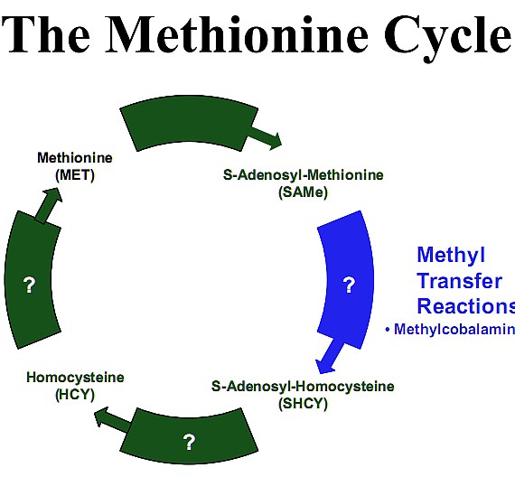
Methionine is converted back into S-Adenosyl-Methionine (SAMe) by giving up its methyl group to a number of different compounds. In this way many types of methylated compounds are produced13.
One of those compounds is N-methylhistamine. The enzyme “Histamine N-methyltransferase” (HMT) catalyzes the transfer of a methyl group from S-Adenosyl-Methionine (SAMe) to the secondary amino group of the imidazole ring of Histamine forming N-methylhistamine14.

So to reiterate and pick up an earlier step, cobalamin assumes two principal forms one of which is methylcobalamin. When hydroxocobalamin is transported into cells via the transcobalamin II transport protein (TCII), this transporter is degraded and hydroxocobalamin converted into 5-deoxyadenosylcobalamin which acts in the mitochondria and methylcobalamin which acts in the cellular cytoplasm. It is there that methylcobalamin is used in the Methionine Cycle to recycle Homocysteine back to Methionine. In this reaction B12 is the cofactor for the enzyme Methionine Synthase (these are the two initiators of this process). Methionine is an essential amino acid that is used to make SAMe. SAMe is the body’s primary methyl donor for methylation reactions. One of those reactions indirectly converts Histamine to N-methylhistamine.
How does converting Histadine to N-methylhistamine increase hunger?
Alterations in brain histidine (and histamine) concentration are associated with changes in food intake. There is an inverse relationship between brain histidine (and histamine) and food intake such that elevated levels reduce hunger and reduced levels increase hunger15.
In addition the central histamine receptors (H1, H2 and H3) are involved in the regulatory process. Antagonizing histamine H1 receptors stimulate appetite and weight gain16-20.
On the other hand, it has been found that cerebroventricular infusion of an H3 receptor antagonist (thereby increasing synthesis and release of brain histamine) reduces hunger and depresses feeding in rats21.
However those compounds that are H3 receptor agonists (they decrease synthesis and release of brain histamine) increase hunger and feeding. N-Methylhistamine is an agonist at H3 receptors22,23.
So it would be expected that decreasing the amount of appetite suppressant (histamine) by converting it to an appetite stimulant N-Methylhistamine would have an overall effect of increasing appetite. This increased hunger effect was discovered more then half a century ago.
References:
1 – Wetzel, Norman C. et al., Growth Failure in School Children as Associated with Vitamin B12 Deficiency—Response to Oral Therapy, Science 16 December 1949 110: 651-653
2 – Lueck, B. W. 1949, The Effect Of Vitamin B12 Concentrate On The Growth Of Weanling Pigs Fed Corn-Soybean Diets AScience, 110: 139
3 – Bosshardt, D. K., W. J. Paul Ande. H. Barnes 1950, The Influence Of Diet Composition On Vitamin B12 Activity In Mice, J. Nutrition , 40: 595
4 – Anderson,G. C., Anda. G. Hooan 1950, Requirement Of The Pig For Vitamin B12, J. Nutrition, 40: 243
5 – Ralli, E., et al., The Effects In Rats Of Vitamin B12, With And Without Ethyl Alcohol, On Nitrogen Balance, Serum Albumin, Liver Nitrogen And Fat, J. Nutr., Jan 1959; 67: 41 – 57
6 – Ershoff, B. H. 1947, Comparative Effects Of Liver And Yeast on Growth And Length Of Survival Of The Immature Thyroid-Fed Rat, Arch. Biochem., 15: 365
7 – Rupp, J., K. E. Paschkisanda. Cantarow 1951, Influence Of Vitamin B12 And Liver Extract On Nitrogen Balance Of Normal And Hyperthyroid Rats, Proc. Soc. Exp. Biol. Med., 76: 432
8 – Wilde, E., The treatment of growth failure in Aleut school children, J. Pediat., 40: 565, 1950
9 – Larcomb, J. W., Perry, C. S., and Peterman, R. A., Dietary supplementation of vitamin B12 in prepuberty school-age children, J. Pediat., 45:70, 1954
10 – COMMITTEE ON NUTRITION: Appraisal of the Use of Vitamins B1 and B12 as Supplements Promoted for the Stimulation of Growth and Appetite in Children, Pediatrics, May 1958; 21: 860 – 864
11 – Schlutz F. W., et al., The Effect Of Varied Vitamin B Ingestion Upon The Appetite Of Children, The Journal of Nutrition, Vol. 15, No. 5 May, 1938
12 – Wheatley, Carmen, A scarlet pimpernel for the resolution of inflammation? The role of supra-therapeutic doses of cobalamin, in the treatment of systemic inflammatory response syndrome (SIRS), sepsis, severe sepsis, and septic or traumatic shock, Medical Hypotheses (2006) 67, 124–142
13 – Springer Handbook of Enzymes Volume 28, Springer Berlin Heidelberg 2006, pages 43-50
14 – Schwelberger HG, Histamine N-methyltransferase (HNMT) enzyme and gene. In Falus A (ed). Histamine: Biology and Medical Aspects, SpringMed Publishing, Budapest, 2004: 53-59
15 – Mercer, L. P., Manipulation of Central Nervous System Histamine or Histaminergic Receptors (H1) Affects Food Intake in Rats, J. Nutr. 124: 1029-1036, 1994
16 – Kalucy, R. S. (1980), Drug-induced weight gain, Drugs 19: 268-278
17 – Kanba, S. & Richelson, E. (1991), Interactions with psychotropic drugs. In: Histaminergic Neurons: Morphology and Function (Watanabe, T. & Wada, H., eds.), pp. 271-282, CRC Press, Boca Raton, FL.
18 – Lavenstein, A. F., Dacaney, E. P., Lasagna, L. &. Van Metre, T. E. (1962), Effect of cyproheptadine on asthmatic children, JAMA 180: 912-916
19 – Noble, R. E. (1969), Effect of cyproheptadine on appetite and weight gain in adults, JAMA 209: 2054-2055
20 – Silverstone, T. & Schuyler, D. (1975), The effect of cyproheptadine on hunger, caloric intake and body weight in man, Psychopharacologia 40: 335-340
21 – Machidori, H., Sakata, T., Yoshimatsu, H., Ookuma, K., Fujimoto, K., Kurokawa, M., Yamatodani, A. & Wada, H. (1992), Zucker obese rats: defect in brain histamine control of feeding, Brain Res. 590: 180
22 – Beales, I. and Calam, J., The histamine H3 receptor agonist N-methylhistamine produced by Helicobacter pylori does not alter somatostatin release from cultured rabbit fundic D-cells, Gut. 1998 August; 43(2): 176–181
23 – Courillon Mallet A, Launay JM, Roucayrol AM, et al., Heli-cobacter pylori infection: physiopathologic implication of N alpha-methyl histamine, Gastroenterology 1995;108:959–66
Anecdotal reports on the effectiveness of long-term supra-physiological administration of Vitamin B12
In 1987 a small town medical clinic witnessed the retirement of it’s general practitioner; a doctor who had practiced by himself in that facility for more then forty years. The clinic was taken over by a new doctor and the case files reviewed. It was discovered that 120 patients had been receiving regular cyanocobalamin (vitamin B12) injections for an average of ten years. Only 4 of those patients however met the current doctor’s criteria for continuing administration. A number of the patients were interviewed to find out their reasons for accepting Vitamin B12 administrations and on a scale of 0 to 4 rated the effectiveness of treatment. The scale was such that 0 represents “no relief”, 1 represents “minimal relief”, 2 represents “fair relief”, 3 represents “good relief” and 4 represents “complete relief”. 1
The patients were then presented with discussion concerning the reason that the doctor felt they did not need vitamin B12. The results of this persuasion were reported in a study which appeared in JAMA with the sole focus of demonstrating that patients could be persuaded to give up vitamin B12. They were able to persuade 62% to give up their injections. 1
The choice of words by the authors of that study reveals their bias when they state, “Anecdotal evidence suggests widespread use of cyanocobalamin as a tonic or as treatment for nonspecific complaints.”
What is more interesting is that 18 patients (38%) who were younger and who reported greater symptom relief refused to give up the Vitamin B12 injections despite the “persuasive evidence” of the new doctor and stated that they would actively seek a physician who would continue to administer cyanocobalamin.1
The most interesting part of the study is the table reproduced below which indicates which ailments the patients had received vitamin B12 for and their perception of effectiveness. Keep in mind that of the 120 patients, 86 were women and 34 were men with a mean age of 71 years. However they had been receiving the administrations for an average of ten years.1
I believe this report is a solid survey of people who are in a position to report the effects of long-term usage. Notice that the largest group of patients received the shots to fight fatigue and weakness. They reported that they had experienced “good relief”.1
It is also noteworthy that in those that used vitamin B12 to increase hunger the effectiveness rating was between good and complete relief.

References:
1 – Lawhorne L MD and Ringdahl, David MD, Cyanocobalamin Injections for Patients Without Documented Deficiency: Reasons for Administration and Patient Responses to Proposed Discontinuation, JAMA 1989;261:1920-1923
Vitamin B12 a Master Molecule
If you are someone who uses vitamin B12 solely to reduce fatigue and increase hunger you may be of the mind that the remainder of this article is not directly applicable to you.
The rest of the article will explore the misdiagnosis of vitamin B12 as a medical failure that may lead to needless suffering in people you care about. Perhaps as a result of what you are about to read you will be in a better position to prompt others around you to demand to be tested properly and to seek accurate diagnosis of their current health problems: Problems that neither they nor their doctors attributed to depletion of this crucial molecule.
In addition the article will touch on vitamin B12’s newly discovered ability to act as an intracellular antioxidant, its role in inflammatory states and the current hypothesis that it is the concentration of vitamin B12 that the body attempts to increase within damaged tissue in disease states. These sorts of things are novel and are directly applicable to everyone irregardless of their current levels.
Deficiency
Absorption of vitamin B12 from food is complex. The vitamin B12 in food is bound to animal protein and must be cleaved via the enzyme pepsin. Pepsin can only be produced in adequate amounts if the stomach contains enough hydrochloric acid. Once isolated B12 is carried to the small intestine by proteins called R-binders. In the intestine, intrinsic factor (IF) which is made in the stomach, attaches to B12 with the assistance of enzymes called pancreatic proteases and carries it to the final section of the small intestine, the ileum. The cells that line the ileum contain receptors that grab the B12-IF complex pulling it into the bloodstream.35

In the bloodstream, another protein, transcobalamin II (TCII) carries vitamin B12 which started as cobalamin but at the stage of entering the blood stream is in the form of Hydroxocobalamin. The carrier protein, Transcobalamin II (TCII) bound to Hydroxocobalamin enters cells in all tissues via the TCII endocytosis ion channel receptor where after TCII degradation it is converted to Methylcobalamin and 5-deoxyadenosylcobalamin and primarily retained intracellularly. A small portion is however exported via the carriers TCII and Transcobalamin III (TCIII). Methylcobalamin acts in the cytoplasm where it is involved in the Methionine Cycle and 5-deoxyadenosylcobalamin in the mitochondria where it is involved in the Krebs cycle (cellular energy).35

This complexity of the vitamin B12 metabolic process greatly exceeds that for any other vitamin and may fail at any one point. The most well known breakdown point, although not the most common is pernicious anemia. This disease occurs when the body fails to produce intrinsic factor (IF) making dietary B12 useless.
Less well-known but more common breakdown points result in lower amounts of vitamin B12 absorption from either food or oral supplementation. Once someone becomes deficient , the few micrograms from standard supplementation will fail to meet the immediate need for thousands of micrograms.
Loss of brain function isn’t always incurable
“Many people do suffer from Alzheimer’s and other dementias but some who are labeled as having dementia are actually suffering from problems that can be corrected”, writes neurologist Sydney Walker III, M.D. Studies suggest that up to 60 percent of patients tentatively labeled as having dementia actually have treatable reversible disorders.1
There is no test that can diagnose Alzheimer’s conclusively in a living patient. The only way to differentiate between Alzheimer’s and other causes of dementia is to rule out all other causes. This is rarely done. In Finland, a study found that only 20 percent of patients with symptoms of dementia were screened for vitamin B12 deficiency.2
Not only is a serum vitamin B12 test too infrequently ordered by physicians it is inadequate. Many seniors with borderline or normal serum B12 levels are severely deficient to the point of developing Alzheimer’s-like dementia. This test gives false positives and negatives and is not sensitive enough. Because of these limitations a Urinary Methylmalonic Acid (MMA)/creatine ratio test should be given. It is an extremely accurate test capable of confirming or ruling out B12 deficiency.3
Dr. Eric Norman the inventor of the test used the MMA test to re-evaluate 299 seniors whose B12 results had tested normal using the standard serum B12 test the year before. The retesting discovered that 2 percent had vitamin B12 deficiency. For thirty million United States elderly, 600,000 new cases per year are going undiagnosed.3
Vitamin B12 deficiency often strikes the nervous system causing damage to the soft fatty material called myelin that surrounds and protects nerve fibers. This damage (akin to a fraying of electrical wires) can cause mysterious and painful neurological problems from numbness, tingling, painful limbs, balance problems, vision loss, incontinence, impotence, memory loss, fuzzy thinking, personality changes, depression and dementia.
This can lead to immune system problems as the body begins to fail to produce sufficient white blood cells. The body becomes more susceptible to infections and viruses. Gastrointestinal problems may occur with the loss of ability to regenerate lining.
Feelings of exhaustion may develop due to anemia when the body is unable to carry sufficient oxygen to cells in the body.
Breakdown in the metabolic pathway (The Methionine Cycle) that detoxifies homocysteine occurs which increases risk of coronary, artery disease, stroke and blood clots.
The lining of the uterus and cervix can become compromised leading to opportune cancers.
The failure to initially diagnose vitamin B12 deficiency and treat it results in a lost opportunity to halt a cascade of problems. Most of these problems will be treated by drugs and therapies none of which attack the root of the problem. With the passage of time eventually remedying the root of the problem with B12 restoration may come too late. The damage that was done will not become undone. There seems to be a critical window of opportunity for treating B12 deficiency and therapy started more than six months after the onset of symptoms may fail to reverse symptoms.
Dr. Mark Goodman reported treating 24 demented patients whose seemingly normal serum B12 levels masked a severe B12 deficiency. When he gave these patients B12 injections every one of them improved dramatically.4
These problems are not confined to the elderly. A twenty-one year old woman who developed bipolar disorder and then full-fledged dementia both due to deficient B12 levels and an accompanying folate deficiency was treated with vitamin B12 and in the words of her doctor experienced a “dramatic resolution” of her symptoms.5
Alzheimer’s
There is some evidence that deficient vitamin B12 levels not only worsen Alzheimer’s symptoms but may possibly play a role in bringing about the disease. The evidence is speculative.
In a study 6 by Robert Clarke levels of B12, folate and homocysteine in 164 of his patients diagnosed with Alzheimer’s were measured and compared to a non-diseased control group. Seventy-six of those patients subsequently died and autopsies confirmed that they did in fact have Alzheimer’s. They found that 6:
- homocysteine levels at the patients’ initial visits were significantly higher in the Alzheimer’s group then the control group;
- B12 and folate levels were significantly lower in the Alzheimer’s group;
- Alzheimer’s patients with high homocysteine levels showed greater evidence of disease progression a trend followed by B12 and folate as well;
- Homocysteine levels in the Alzheimer’s patients didn’t change as the disease progressed indicating that the differences between patients and control group members weren’t caused by the disease itself, but rather predated or coincided with the onset of the disease
He speculated that high levels of toxic homocysteine possibly resulting from B12 deficiency, may cause microinfarcts (tiny areas of blood vessel damage) that then trigger the formation of the plaques and tangles that eventually clutter the brain of a person with Alzheimer’s.6 Robert Clark then concluded “low blood levels of folate and vitamin B12 and elevated total homocysteine levels were associated with Alzheimer’s disease.” 6
In another study demonstrating correlation, researchers found that four of six family members with confirmed Alzheimer’s had low blood levels of B12 while only one of twelve without Alzheimer’s had low levels of B12.7
In still another study demonstrating correlation, researchers collected blood samples from patients before they developed the disease. These samples were compared to samples after they developed the disease. They found that seniors with low intakes of vitamin B12 and folate were twice as likely to develop Alzheimer’s as people with healthy B12 levels.8
In one more study researchers found that Alzheimer’s patients with lower-than-normal B12 levels exhibited more behavioral and psychological symptoms of dementia than patients with normal B12 levels prompting them to conclude “Vitamin B12 could play a role in the pathologenesis of behavioral changes in Alzheimer’s disease.” 9
Multiple Sclerosis and Neurological Disorders
Inadequate levels of vitamin B12 may be misdiagnosed as Multiple Sclerosis or other neurologic diseases in part because the symptoms of B12 deficiency may be identical to neurological disease symptoms. Doctors usually look for blood abnormalities when they check for B12 deficiency however neurological damage can precede blood anomalies by years or even decades. B12 deficiency is common, has many causes and may be found in people of all ages. Yet most doctors believe it to be rare or something that happens in old age. As a consequence whether a patient suffers permanent neurological damage or recovers from inadequate B12 hinges on whether his attending doctor is well informed.
Here are two cases reported in the literature that contrast and dramatize the difference.
Informed Doctor = recovery:
Mystified doctors sent a twenty-eight year old woman to physician Helmut Wilhelm after she developed unexplained vision loss. In addition to checking the woman for other common causes of sudden blindness, Wilhelm found that her serum B12 levels were one-third of the normal limit. he started vitamin B12 injections immediately leading to “an almost complete recovery.” 10
Uninformed and arrogant Doctor = permanent damage:
John Hotchkiss, Jr. MD. practiced in a medical group. One of his partners, a Harvard graduate objected strenuously to his insistence on ruling out B12 deficiency in patients with neurological disorders or other suspicious signs and symptoms. The Harvard doctor grew hostile on more than one occasion when Hotchkiss suggested evaluating a patient’s B12 levels and he refused to be swayed by the medical literature Hotchkiss sent him.
Some years later, Hotchkiss recounted “he sent a patient to me for dizziness evaluation.” Hotchkiss discovered that the woman was not dizzy but instead suffered from poor coordination stemming from neurological dysfunction. He wasn’t surprised because the woman’s history included a gastrectomy 12 years earlier, a surgery that inevitably results in severe B12 deficiency if patients don’t receive compensatory B12 injections.
The woman had been followed by Hotchkiss’ Harvard colleague ever since the surgery, Hotchkiss recounts, “but he had done no follow-up studies concerning her B12 status.” As a result, she suffered from a condition known as “combined systems disease” a complication of chronic B12 deficiency in which damage to the spinal cord tracts causes irreversible crippling.
“The doctor not only failed to monitor her and prevent this from happening, a grave dereliction but he failed to recognize the condition when it occurred”, Hotchkiss recounts. “When we met in the hall some weeks later he said, ‘You got me’.” 11
It has been estimated that up to ten percent of those diagnosed with multiple sclerosis don’t actually have the disease. 12 Most of the time if doctors rule out B12 deficiency in patients suspected of multiple sclerosis they do so by relying on a serum B12 test. This can be inaccurate. Most fail to order other tests such as the urinary MMA test and the plasma Homocysteine test.
Experts (as opposed to doctors in general) believe that a serum B12 level below 350 pg/ml warrants further testing without regard for the wider reference ranges. The urinary MMA should be 3.8ug MMA/mg creatine or above. If not then the patient is likely deficient and a plasma homocysteine test will establish if homocysteine is within the normal range of 4.0 – 12.0 umol/L.13,14
Dr. Eric Norman reported in 2000 that of six women his research team diagnosed with B12 deficiency, three were initially suspected of having multiple sclerosis (MS). These three women’s original doctors were puzzled and diagnosis was delayed because of the similarities between B12 deficiency and MS and because they were young. Dr. Norman commented, “This population deserves further evaluation since it has not been considered prone to cobalamin deficiency.” Proper treatment of his six patients resulted in a nearly total recovery for two and a partial recovery in another. The extent of improvement in two other women could not be measured since treatment had just begun when the report was published and the final woman had not yet developed significant neurologic symptoms. 15
Cases reported in the literature reveal that this is not an isolated case. Another example of B12 deficiency diagnosed as MS was reported in the American Journal of Psychiatry by Dr. Gary Payinda.16
At fifty-two years of age, Mrs. A suddenly developed paralysis in her legs. Her doctor referred her to a neurologist, who diagnosed her with multiple sclerosis. Over the next two months, Mrs. A was placed on numerous medications, but her leg weakness progressed and the drugs did not help. She required a cane, then used a walker and eventually needed a wheelchair to get around. As time went by, Mrs. A became agitated and angry. She grew paranoid and she called the police to report that her family was trying to poison her. She also became violent, throwing furniture and even trying to jump from a moving car.
Mrs. A’s family stunned and frightened by her worsening behavior finally took her to an emergency psychiatric center. She appeared disheveled was delirious and disoriented and paranoid and could not stand without assistance. The psychiatric facility obtained a B12 level which came back extremely low at 9pg/ml.
The doctors diagnosed Mrs. A. with subacute combined spinal cord degeneration and psychosis due to severe vitamin B12 deficiency. Additional tests revealed that she suffered from pernicious anemia; earlier physicians missed the diagnosis in part because her folic acid supplements had masked her blood abnormalities.
Two days after starting B12 injections, Mrs. A. started regaining the strength in her legs. Within eight weeks, her symptoms of mental illness vanished. Unfortunately she may never fully regain her health and mobility because of the delay in diagnosing her correctly. The cause of that delay: her original neurologist failed to diagnose her correctly, instead misdiagnosing her with MS and apparently never considering B12 deficiency.16
Depression
Vitamin B12 deficiency attacks nerves by stripping them of their protective myelin coating and disrupting the communication between brain cells and the nervous system. While this can create the types of symptoms associated with multiple sclerosis it can also affect the nerve cells in the brain that control how one feels, thinks and behaves. Deficiency can cause severe mental illness, including depression, paranoia and even symptoms that resemble schizophrenia.
A few examples will serve to underscore the link between B12 and depression.
Researchers at the National Institute on Aging evaluated a group of disabled women over the age of sixty-five who were active and living independent lives and found that B12 deficiency doubled the risk of severe depression in that group. They concluded that, “It should be an alarming sign that we found a significant rate of B12-deficiency caused depression in this population.”17
In a very comprehensive Netherlands study, researchers screened almost four thousand older individuals for depression and then compared the laboratory test results of those with depressive symptoms to non-depressed control group members. The researchers report that high homocysteine levels, vitamin B12 deficiency and to a lesser extent folate deficiency were all related to depressive disorders. When they controlled for other factors, the effects of homocysteine and folic acid levels were less prominent but low B12 levels were still strongly associated with depression. 18
Here are a few case studies from medical journals:
Doctor G. Daynes reported that in his own practice as medical director of a hospital in South Africa, he successfully treated eight women whose postpartum psychosis stemmed from B12 deficiency. His patients’ recoveries led him to recommend that all women with postpartum psychosis receive large doses of B12. “Where the postpartum psychosis is not primarily caused by lack of vitamin B12, the giving of the preparation will do no harm,” he remarked, “so it seems to me that in all such cases it should be given as soon as possible.”19
Doctors treating a twenty-year old woman, who had attempted suicide three months earlier, discovered the reason for the woman’s urges to kill herself. She had autoimmune pernicious anemia. She now receives regular injections of B12, which have eliminated her depression and suicidal thoughts.20
Dr. Frederick Goggans and colleagues reported a case of an elderly man who suddenly developed severe mania, believing that his hometown was planning a large celebration in his honor, including appearances by Hollywood celebrities. “He became so physically energized,” the doctors wrote, “that six younger men were required to restrain him at the time of admission to the hospital.” Lab tests demonstrated low B12 level and his doctors eventually diagnosed pernicious anemia and treated him with B12 injections. He recovered quickly and at his six-month checkup he as doing well.21
Cardiovascular Problems: B12 – Homocysteine
When vitamin B12 levels drop too low the Methionine Cycle breaks down which results in elevated levels of homocysteine. That cycle converts methionine into smaller molecules one of which is SAMe. SAMe then further breaks down into thousands of compounds and proteins that are vital for healthy cells, tissue and organs. One of those breakdown products is homocysteine. A properly functioning cycle results in homocysteine getting quickly recycled back into methionine with the assistance of vitamin B12 and folic acid. Although there are other pathways for rendering homocysteine harmless, if someone is deficient in B12 the normal functioning of the primary pathway the Methionine Cycle is disrupted. This results in excess homocysteine.
Excess amounts cause blood vessels to lose their elasticity, making it harder for them to dilate and damaging the inner lining. This damage allows cholesterol, collagen and calcium to attach to the inner walls of the blood vessels where they can form sticky deposits called atherosclerotic plaque. These plaques narrow arteries and increase the risk of artery disease, heart attacks, strokes, blood clots and aneurysms.22
Homocysteine is also an oxidant that decreases the production of nitric oxide, a substance crucial to healthy blood vessel function. Decreased levels of nitric oxide are linked to atherosclerosis and high blood pressure.
Homocysteine has been shown to precede the onset of cardiovascular disease.23 Five to ten percent of the population and as many as 30 to 40 percent of senior citizens have high homocysteine levels.24
Link between homocysteine and cardiovascular disease
Researchers in Israel found that male children of heart attack victims had significantly higher homocysteine levels then control group members.25 Furthermore they found that the male children in the top 20 percent were far more likely than others to be in the “parental heart attack” group.
The researchers noted that men in Jerusalem had much higher homocysteine levels then men in the United States which explained the higher heart attack rate. They then added that the difference in homocysteine levels between men in the two countries “was largely attributable to lower plasma vitamin B12 levels in the Israeli population.”25
In a dramatic study reported in the New England Journal of Medicine, Norwegian researchers followed 587 patients with existing coronary artery disease. Within five years of undergoing treatment 11 percent of the patients had died. ‘We found a strong, graded relation between plasma homocysteine levels and overall mortality,” the researchers reported, with about 4 percent of the patients in the low-homocysteine group dying as compared to nearly 25 percent of those in the high-homocysteine group.22 Similar results were found in another study following 400 patients who suffered heart attacks. They discovered that the long-term death rate was more than twice as high in the upper 40 percent of homocysteine levels as it was for other patients.26
More evidence comes from a meta-analysis study which pooled data from seventy-two studies. They concluded that there is “strong evidence that the association between homocysteine and cardiovascular disease is causual”. 27
In young people as well research has demonstrated that high homocysteine is a threat. A large-scale study found that elevated homocysteine nearly doubles the risk of stroke in women between the ages of fifteen and forty-four. The researchers concluded, “The magnitude of the increase in stroke risk was similar to that of smoking a pack of cigarettes per day.”28
B12 lowers homocysteine
Another study, this one short-term (only eight weeks) tested the effects of folic acid alone, B12 alone, and folic acid plus B12 in lowering the homocysteine levels of patients who had suffered ischemic strokes. The researchers found that all three approaches worked but that “the combination therapy yielded the most remarkable result, i.e. plasma total homocysteine was reduced by 38.5 percent.29 A similar short-term study found a 23 percent decrease in homocysteine levels after six weeks of treatment with folic acid and B12.30
Lowering homocysteine reduces cardiovascular problems
Researchers in Switzerland offered homocysteine-lowering therapy to half of a group of 553 patients who had undergone angioplasty to correct coronary artery stenosis (narrowing). After one year, they found that the incidence of “major adverse events” – deaths, nonfatal heart attacks, or the need for repeat angioplasty – was 33 percent lower in the treatment group.31
In another study, the same Swiss research group administered folic acid, B12 and B6 or a placebo to 205 patients who had undergone successful coronary artery angioplasty. They reported that the rate of restenosis (renarrowing of the arteries) was significantly lower in the treatment group than in the placebo group (19.6 percent versus 37.6 percent) and that this group had less than half the need for a repeated procedure. They concluded, “This inexpensive treatment, which has minimal side effects, should be considered as adjunctive therapy for patients undergoing coronary angioplasty.”32
Finally in a study that evaluated eighty-nine men ranging in age from thirty-nine to sixty-seven with existing coronary artery disease, eight weeks of treatment with folic acid and vitamin B12 significantly reduced plasma homocysteine levels compared to controls. Of interest is that they found that the arteries of men taking the vitamins dilated more efficiently in response to blood flow demands. The researchers concluded, “these findings support the view that lowering homocysteine, through B vitamin supplementation may reduce cardiovascular risk.” 33
Even a major pharmaceutical company had at one time no difficulty admitting all of this. A few years ago on Abbott Laboratories Malaysian website they had a section entitled “Homocysteine: The newly proven risk factor for heart attacks”.
On that page were the following sentences:
“Elevated levels of homocysteine might result from lack of certain vitamins which are involved in the breakdown of homocysteine. The vitamins involved are vitamin B6, vitamin B12 & Folic Acid.” 34
“Homocysteine can be easily controlled by providing our body with Folic acid, Vitamin B6 & Vitamin B12 at amounts exceeding the recommended daily allowances.”34
That page is currently only available through the “Internet Archive Wayback machine.”
Inflammation
Let’s reiterate a part of the vitamin B12 uptake process. In the bloodstream, another protein, transcobalamin II (TCII) carries vitamin B12 which started as cobalamin but at the stage of entering the blood stream is in the form of Hydroxocobalamin. The carrier protein, Transcobalamin II (TCII) bound to Hydroxocobalamin enters cells in all tissues via the TCII endocytosis ion channel receptor where after TCII degradation it is converted to Methylcobalamin and 5-deoxyadenosylcobalamin and primarily retained intracellularly. A small portion is however exported via the carriers TCII and Transcobalamin III (TCIII). Methylcobalamin acts in the cytoplasm where it is involved in the Methionine Cycle and 5-deoxyadenosylcobalamin in the mitochondria where it is involved in the Krebs cycle (cellular energy).35
What is of extreme interest is the well-established observation that cobalamin carrier proteins, the Transcobalamins (TCS) are elevated during trauma, infections and chronic inflammatory conditions. This remains to be explained. It has been proposed that such TC elevations signal a need for cobalamin to resolve the inflammatory event.36
Chronic inflammation is usually accompanied by oxidative stress. These factors are primary components of many age-related diseases including cancer, atherosclerosis, neurodegenerative disease and arthritis. Recent studies suggest that cobalamins (vitamin B12 derivatives) may modulate the oxidative stress responses, including those of the inflammatory response. 36
Those studies specifically found:
- Inflammatory diseases are associated with elevated blood levels of transcobalamin (a cobalamin transport protein) 38; and
- It’s (transcobalamin) membrane receptor is up-regulated by TNF-a 39; and
- Cobalamin concentration also modulates TNF-a levels in cerebrospinal fluid. 40
“TNF-a is important in inflammatory responses, so taken together, these observations suggest that elevation of cobalamin could be used to supplement the cellular response to inflammation.”36
There is also an increase in the unsaturated B12 binding capacity.45,46 All of these events seem to occur in chronic inflammation states such as diabetes, Crohn’s disease, rheumatoid arthritis, systemic lupus erythematosus and acute inflammations such as some cancer states, trauma and infections. The increase in transcobalamin carrier proteins and their binding capacity also increase in B12 deficiency. All of this is indicative of a “crying out” for vitamin B12.
In order to appreciate how B12 relates to inflammation let us expand upon the functions we have previously described herein.
In the cell, two enzymes use cobalamin as a cofactor in either the 5-deoxyadenosylcobalamin(AdoCbl) or the methylcobalamin (MeCbl) form.
In the mitochondria AdoCbl is used by the enzyme L-methylmalonyl-CoA mutase in a reaction whereby L-methylmalonyl-CoA is converted to succinyl-CoA, which then enters the Krebs cycle (cellular energy).
In the cytoplasm methylcobalamin (MeCbl) is used by the enzyme methionine synthase in a reaction, (i.e. The Methionine Cycle) whereby tetra-hydrofolate and methionine are generated by a methyl group transfer from methyltetrahydrofolate to homocysteine.41
Homocysteine is a junction metabolite. This means it can be recycled back into the methionine cycle as described above or it can be converted to cysteine via the transsulfuration pathway, ultimately leading to synthesis of the important intracellular antioxidant glutathione. 41
So when the Methionine Cycle is operating optimally the antioxidant glutathione is synthesized and homocysteine does not become elevated as it is fully occupied in either of its two roles.
However elevated homocysteine is associated with endothelial cell (thin layer of cells that line the interior surface of blood vessels) dysfunction. High levels of homocysteine promote the formation of reactive oxygen species (small molecules that are unstable and highly reactive able to create significant cellular damage), primarily by a mechanism involving endothelial nitric oxide synthase (catalyze the production of nitric oxide in blood vessels). 42
Homocysteine also inhibits the antioxidant enzymes superoxide dismutase (important antioxidant defender) and glutathione peroxidase (main biological role is to protect the organism from oxidative damage) and activates endothelial (blood vessel lining) pro-inflammatory signaling pathways. 43
Elevated Homocysteine levels induce cell death in large part because of the inhibition of glutathione. Reduced glutathione levels have been found to correlate with increased oxidative stress, mitochondrial damage, and apoptosis.44 So when it was very recently discovered that cobalamins protect against cell death induced by homocysteine37 part of that beneficial effect was attributed to cobalamin’s ability to increase intracellular clearance of homocysteine through its role in the Methionine Cycle.
However in that study cobalamins were very protective against damage from the reactive oxidative species, Hydrogen peroxide which indicates that they also act as antioxidants via other mechanisms. 37
The recent Birch study mentioned above was highly significant because it found that cobalamins (specifically thiolatocobalamin) inhibited intracellular peroxide production, maintained intracellular glutathione levels, and prevented apoptotic and necrotic cell death at supraphysiological concentrations. Further these supraphysiological concentrations were not toxic prompting the study to conclude that cobalamins are “powerful but benign antioxidants at pharmacological concentrations.” 37
The study explained their results as follows:
“The antioxidant properties of cobalamin probably result from a combination of direct and indirect effects: stimulation of methionine synthase activity, direct reaction with reactive oxygen and nitrogen species, a glutathione sparing effect, and modification of signaling molecules, leading to induction of stress responses. The remarkably superior protection of thiolatocobalamins in vitro presumably relates to their enhanced function in one or more of these potential mechanisms, the balance of which may differ between the two compounds. In conclusion, cobalamins, and in particular the thiolatocobalamins, exhibit a marked antioxidant activity at pharmacological concentrations and afford significant cellular protection against oxidative stress.”37
Toxicity
Cobalamin is a water-soluble vitamin with a high renal excretion rate and a very safe toxicity profile. Serum levels have been kept as high as 10,000 mcg/ml in infants and children for the long-term treatment of congenital TCII deficiency.47
Hydroxycobalamin has been routinely used in the ICU, in France, and elsewhere, for over 40 years in 4/5 gram iv doses given on up to three consecutive days, as a treatment for cyanide poisoning.48-54 The FDA has granted Hydroxycobalamin orphan drug status for this purpose.54
High dose cobalamin has been used in children, in a 1950s pre-chemotherapy era trial for neuroblastoma where 1000 mcg were given, every other day, for up to 8 years.55
No where in any of these reports is there any indication that high doses led to toxicity. To the contrary, high dose cobalamin is deemed safe.
Cancer
There are certain disease states that result in an elevation in levels of serum cobalamin. Many liver diseases such as hepatitis, cirrhosis and liver cancers as well as leukemias are all accompanied by an increase in circulating cobalamin. This phenomenon is caused predominantly by cobalamin release during what is called “hepatic cytolysis”. Cytolysis is a bursting of the cell and therefore it is not surprising that its contents will be elevated in plasma. Another explanation is that when the liver is in distress there is a decrease in the clearance of compounds, including cobalamin.56
Because of this strong correlation between B12 and cancers you come across studies that find such things as a relationship between vitamin B12 levels and survival in terminally ill cancer patients. In one such study it was found that the length of survival decreased with the increase in serum vitamin B12 levels. The study’s data indicated that an “elevated serum vitamin B12 level is a predictive factor for mortality in patients with cancer.”57
This type of correlation has even led one study58 to draw the erroneous causative conclusion and underscores the need for a determined researcher to discriminate between the conclusions drawn in studies, treating each not as dispositive but rather simply part of an overall picture spanning many multiple disciplines. A researcher that is able to read and draw from a wider array of material is in a better position to come to an understanding.
Vitamin B12 may be elevated in plasma during times of cancer but it is not stimulatory of cancer. The effect of cobalamin on the proliferation of a variety of malignant cells has been examined in vivo and in vitro in numerous studies.59-61 Here are a few very brief examples:
- Methylcobalamin was found to inhibit the proliferation of androgen-sensitive SC-3 cells (a cloned cell line from Shionogi mouse mammary tumor, SC115) in culture at the concentration of 100–300 ug/ml; and
- An inhibitory activity of methylcobalamin on proliferation was also observed in other cell lines (estrogen-sensitive B-1F cells from mouse Leydig cell tumor and MCF-7 cells from human mammary tumor) at the concentration of 500 ug/ml; and
- Large doses of methylcobalamin injected intraperitoneally (100 mg/kg body weight/day) were non-toxic and suppressed the tumor growth of SC115 and B-1F cells in mice fed a vitamin B12 deficient diet.
Vitamin B12 does not in any way stimulate cancer cells. There are no experimental results published indicating that vitamin B12 stimulates growth of malignant cells. In fact these results indicate to the contrary that methylcobalamin inhibits the proliferation of malignant cells in culture and in vivo. A reading of those studies and others find that often the researchers propose the possibility of methylcobalamin as a potentially useful therapeutic agent for the treatment for some malignant tumors.61
B12 Deficiency and Cancer
One of the strongest scientifically emerging associations is one between breast cancer and B12 deficiency. In a Johns Hopkins study, researchers measured the B12 in blood samples taken from women who had donated blood, comparing samples from 195 women who later developed breast cancer to samples from 195 cancer-free women. Among postmenopausal women, the researchers found those whose B12 levels fell into the lowest fifth were two to four times more likely to develop breast cancer than those in the upper four fifths.67
If you have read this far and hadn’t yet identified a reason to supplement with vitamin B12 for either yourself or people you care about, I believe you have just found one.
In addition to breast cancer, other cancers are being tentatively linked by studies to deficient levels of vitamin B12. Among these are cervical, lung and oral cancers.68-72
One explanation for why deficient B12 levels appear to promote the development of cancer is that the body needs ample B12 in order for folate to work. One of folate’s crucial roles is the synthesis of the nucleotide “building blocks” of DNA. When folate is trapped in unusable form due to a lack of B12 it can not perform this role. This leads to an imbalance in the supply of DNA building blocks, forcing the body to make changes in DNA structure that can make the DNA more vulnerable to breakage. Broken DNA strands can lead to mutations that in turn can lead to cancer. Research demonstrates that chromosome breakage is strongly correlated with deficiencies of either folate or B12 (or with high levels of homocysteine, linked to B vitamin deficiencies) and that large dietary supplements of B12 can minimize this breakage.73
Master Molecule
We have barely scratched the surface and in no way have we even begun to examine the details behind the hypothesis that vitamin B12 is a master molecule which regulates the transcription factor, NFxB thereby determining the extent of the inflammatory response and its subsequent resolution. NFxB is the brain’s master regulator switch in inflammation. Cobalamin (B12) appears to regulate NFxB in both its pro and anti-inflammatory roles. The cascade through which this occurs is simply too complex to be discussed in this paper. To give a quick (but very incomplete) feel for the verity of the hypothesis I will point to a model.
Indirect evidence for Cobalamin/NFxB regulation comes from a model of chronic inflammation. Rheumatoid Arthritis has been treated with some success using high doses of Methycobalamin, one of the active forms of cobalamin, to which Hydroxycobalamin is partially converted intra cellularly.62 An in vitro study to determine cytokine production of monocytes exposed to three mitogens or recombinant Interleukin-2, found Methycobalamin suppressed levels of Interleukin-6 production with three mitogen-stimulation by 66%, 68% and 81%, as compared to controls. Suppression of Interleukin-6 by Methylcobalamin was dose-dependent, and not total.62 Interleukin-6 is believed to be an accurate marker for TNFa activity.63 Both TNFa and Interleukin-6 are pro-inflammatory cytokines (signaling substances) and under run-away inflammatory events such as severe sepsis, and septic or traumatic shock signal the release of still more inflammatory agents. Suppressing their production during periods of inflammation is a sign that Vitamin B12 is regulating NFxB (the inflammation switch).
For a much fuller, detailed look at this hypothesis, three papers by Carmen Wheatley cited below should be read.64-66
Conclusion
You can take steps to lower excess triglycerides, LDL and cholesterol, raise HDL. You can take all the fish oil you want, and modify your diet to lower glucose and reduce insulin. You can modify your lifestyle to avoid hypertension and seek out hormone replacement therapy. But if your levels of homocysteine are excessive all of your changes were for naught. You will suffer from adverse cardiovascular consequences.
Vitamin B12 is a molecule that is vital to maintaining a healthy body. It doesn’t matter whether it proves to be a master molecule. Its presence maintains health and its absence leads to degeneration. It is a molecule that is not toxic if taken in excess. Waiting for the symptoms of malady to appear before introducing vitamin B12 to a regular supplementation protocol may engender deficits that can not be fully overcome. Adding vitamin B12 to a supplementation protocol designed to maintain health and vigor may avoid many of the potentialities discussed herein. Vitamin B12 is by no means a panacea but neither is it snake oil. It is simply a vital molecule of which four out of every ten people are lacking.
Click Here to Purchase Synthelamin!
References:
1 – Walker III, Sydney, Dose of Sanity, New York: John Wiley & Sons, 1996 p. 192
2 – Lopponen, M., et al., Diagnosing cognitive impairment and dementia in primary health care – a more active approach is needed, Age and Ageing, 32(6):606-12
3 – Norman, E.J., Vitamin B12 deficiency, Journal of Family Practice (1993), 36:597
4 – Interview with mark Goodman in Clinical Pearls News (1997), 7(10):132-134
5 – Reid, S.D., Pseudodementia in a twenty-one year old with bipolar disorder and vitamin B12 and folate deficiency, West Indian Medical Journal (2000), 49(4):347-8
6 – Clarke R., et al., Folate, vitamin B12 and serum total homocysteine levels in confirmed Alzheimer’s disease, Archives of Neurology (1998), 55:1449-55
7 – McCaddon, A., Familial Alzheimer’s disease and vitamin B12 deficiency, Age and Ageing (1994), 23(4):335-7
8 – Wang, HX. et al., Vitamin B12 and folate in relation to the development of Alzheimer’s disease, Neurology 2001, 56:1188-94
9 – Meins, W. et al., Subnormal serum vitamin B12 and behavioural and psychological symptoms in Alzheimer’s disease, International Journal of Geriatric Psychiatry (2000), 15(5):415-8
10 – Wilhelm, H., Grodd, W., Scheifer, U. and Zrenner, E., Uncommon chiasmal lesions: demyelinating disease, vasculitis and cobalamin deficiency, German Journal of Ophthalmology 1993, 2:234-40
11 – Hotchkiss, J., A Controversial Vitamin (conference presentation), June 2001, Society for Orthomolecular Medicine, San Francisco
12 – Physician information posted on the Neurology and neurosurgery Forum in answer to a question. Questions answered by physicians from the Clevaland Clinic
13 – The writings and reports of Sally M. Pacholok, R.N. (a studied expert on the topic of Vitamin B12 Misdiagnoses)
14 – Goodikin, D.E. et al., Serum cobalamin deficiency is uncommon in multiple sclerosis, Internal Medicine 1994, 33:82-86
15 – Norman, E. J., Cobalamin (vitamin B12) deficiency identified in young Caucasion women, Blood, 2000 96(11):8b
16 – Payinda, G. and Hansen, T., Vitamin B(12) deficiency manifested as psychosis without anemia, America Journal of Psychiatry 2000, 157:660-61
17 – Penninx, B.W., et al., Vitamin B12 deficiency and depression in physically disabled older women: Epidemiologic evience from the Women’s Health and Aging Study, American Journal of Psychiatry 2000 May;157(5);715-21
18 – Tiemeier, H., et al., Vitamin B12, folate and homocysteine in depression: The Rotterdam Study, American Journal of Psychiatry 2002, 159(12):2099-101
19 – Daynes, G. , Cyanocobalamin in postpartum psychosis, South Africa Medical Journal 1975, 49(34):1373
20 – Middleman, A. B., et al., A routine CBC leads to a non-routine diagnosis, Adolescent Medicine 1996, 7(3):423-6
21 – Goggans, F.C., A case of mania secondary to vitamin B12 deficiency, American Journal of Psychiatry 1984 141(2):300-1
22 – Nygard, O. et al., Plasma homocysteine levels and mortality in patients with coronary artery disease, New England Journal of medecine 1997, 337:230-6
23 – O’Callaghan P., et al., Update on homocysteine, Heartwise (Irish heart Foundation), Winter 2000
24 – Booth, G. L. and Wang, E. E., Preventative halth care 2000 update: Screening and management of hyperhomocysteinemia for the prevention of coronary artery disease events, The Canadian Task Force on Preventative Health care, Canadian medical Association Journal 2000, 163(1):21-9
25 – Kark, J.D., et al., Plasma homocysteine and parental myocardial infarction in young adults in Jerusalem, Circulation 2002, 105(23):2725-9
26 – Stubbs, P.J., et al., Effect of plasma homocysteine concentration on early and late events in patients with acute coronary syndromes, Circulation 2000, 102:605-10
27 – Wald, D.S., et al., Homocysteine and cardivascular disease: Evidence on causality from a meta-analysis, British Medical Journal 2002, 325:1202
28 – Kittner, S.J., et al., Homocyst(e)ine and risk of cerebral infarction in a biracial population: The stroke prevention in young women study, Stroke 1999, 30(8):1554-60
29 – Sato, Y., et al., Hyperhomocysteinemia in Japanese patients with convalescent stage ischemic stroke: Effect of combined therapy with folic acid and mecobalamine, Journal of the Neurological Sciences 2002, 202(1-2):65-8
30 – MacMahon, M., et al., Nutrition, Metabolism, and Cardiovascular Diseases 2000, 10(4):195-203
31 – Schnyder, G., et al., Effect of homocysteine-lowering therapy with folic acid, vitamin B(12), and vitamin B(6) on clinical outcome after percutaneous cornary intervention, The Swiss Heart Study: a randomized controlled trial, Journal of the American Medical Association 2002, 288(8):973-9
32 – Schnyder, G., et al., Decreased rate of coronary restenosis after lowering of plasma homocysteine levels, New England Journal of Medicine 2001, 345(22):1593-600
33 – Chambers, J.C., et al., Improved vascular endothelial function after oral B vitamins. An effect mediated through reduced concentrations of free plasma homocysteine, Circulation 2000, 102:2479-83
34 – https://www.abbott.com.my/t_healthv_main.html
35 – Nexo, E., Cobalamin binding proteins. In: Krautler B., et al., Vitamin B12 and B12 proteins, La Jolla: Wiley; 1998 p. 461-75
36 – Wheatley, Carmen, A scarlet pimpernel for the resolution of inflammation? The role of supra-therapeutic doses of cobalamin, in the treatment of systemic inflammatory response syndrome (SIRS), sepsis, severe sepsis, and septic or traumatic shock, Medical Hypotheses (2006) 67, 124–142
37 – Birch, C. S., et al., A novel role for vitamin B12: Cobalamins are intracellular antioxidants in vitro, Free Radical Biology & Medicine 47 (May 2009) 184-188
38 – Rothenberg, S.; Quadros, E.; Regec, A., Transcobalamin II, In Chemistry and Biochemistry of B12 (ed R. Banerjee) pp441–473. Wiley, New York; 1999
39 – Kalra, S.; Ahuja, R.;Mutti, E.; Veber, D.; Seetharam, S.; Scalabrino, G.; Seetharam, B., Cobalamin-mediated regulation of transcobalamin receptor levels in rat organs, Arch. Biochem. Biophys. 463:128–132; 2007
40 – Scalabrino, G.; Carpo, M.; Bamonti, F.; Pizzinelli, S.; D’Avino, C.; Bresolin, N.; Meucci, G.; Martinelli, V.; Comi, G. C.; Peracchi, M., High tumor necrosis factor-alpha levels in cerebrospinal fluid of cobalamin-deficient patients, Ann. Neurol. 56: 886–890; 2004
41 – McCaddon, A.; Regland, B.; Hudson, P.; Davies, G., Functional vitamin B12 deficiency and Alzheimer disease, Neurology 58:1395–1399; 2002
42 – Ginnan, R.; Guikema, B. J.; Halligan, K. E.; Singer, H. A.; Jourd’heuil, D., Regulation of smooth muscle by inducible nitric oxide synthase and NADPH oxidase in vascular proliferative diseases, Free Radic. Biol. Med. 44:1232–1245; 2008
43 – Biswas, S.; Chida, A. S.; Rahman, I., Redoxmodifications of protein-thiols: emerging roles in cell signaling, Biochem. Pharmacol. 71:551–564; 2006
44 – Vitvisky, V.;Mosharov, E.; Tritt, M.; Ataullakanov, F.; Banerjee, R., Redox regulation of homocysteine-dependent glutathione synthesis, Redox Rep. 8:57–63; 2003
45 – Olesen H, Andersen MP, Amris A., Serum vitamin B12 binding capacity in patients with anaemia, Scand J Haematol 1968;5:235–40
46 – Ghosh K, Mohanty D, Rana KS, et al., Plasma transcobal- amins in haematological disorders, Folia Haematol Int Mag Klin Morphol Blutforsch 1986;113:766–75
47 – Fenton WA, Rosenberg LE., Inherited disorders of cobalamin transport and metabolism. In: Scriver CR, Badet AL, Sly WS, Valle D, editors. The metabolic basis of inherited disease, New York: McGraw-Hill; 1995. p. 3129–50
48 – Yacoub M, Faure H, Morena M, et al., L‘intoxication cyanhydrique aigue Donnees actuelles sur le metabolisme du cyanure et le traitement par hydroxocobalamine, J Europ de Toxicol 1974;7:22–9 [In French]
49 – Motin J, Bouletrean P, Rouzious JM., Intoxication cyanhydrique grave traitee avec succes par hydroxocobalamine, J Med de Strasbourg 1970;I:717–22 [In French]
50 – Tassan H, Joyon D, Richard T, et al., Intoxication au cyanure de potassium traitee par l’hydroxocobalamine, Ann Fr Anesth Reanim 1990;9:383–5 [In French]
51 – Brouard A, Blaisot B, Bismuth C., Hydroxocobalamine in cyanide poisoning, J de Toxicol Clin et Exp 1987;7:155–68
52 – Forsyth JC, Mueller PD, Becker CE, et al., Hydroxocobalamin as a cyanide antidote: safety, efficacy and pharmacokinetics in heavily smoking normal volunteers, Clin Toxicol 1993;31:277–94
53 – Riou B, Gerard JL, Drieu la Rochelle C, et al., Hemodynamic effects of hydroxocobalamin in conscious dogs, Anaesthes 1991;74:552–8
54 – Hall AH, Rumack BH., Hydroxocobalamin/sodium thiosulfate as a cyanide antidote, J Emerg Med 1987;5:115–21
55 – Bodian M., Neuroblastoma. Pediatr Clin North Am 1959;6:449–72
56 – Ermens AA, Vlasveld LT, van Marion-Kievit JA, Lensen CJ, Lindemans J., The significance of an elevated cobalamin concentration in the blood, Ned Tijdschr Geneeskd 2002;146(10):459–64
57 – Geissbuhler P, Mermillod B, Rapin CH., Elevated serum vitamin B12 levels associated with CRP as a predictive factor of mortality in palliative care cancer patients: a prospective study over five years, J Pain Symptom Manage 2000;20(2):93–103
58 – Hultdin J, Van Guelpen B, Bergh A, Hallmans G, Stattin P., Plasma folate, vitamin B12, and homocysteine and prostate cancer risk: a prospective study, Int J Cancer 2005;113(5): 819–24
59 – Nishizawa Y, Yamamoto T, Terada N, Fushiki S, Matsumoto K, Nishizawa Y., Effects of methylcobalamin on the proliferation of androgen-sensitive or estrogen-sensitive malignant cells in culture and in vivo Int J Vitam Nutr Res 1997;67(3):164–70
60 – Liteplo RG, Hipwell SE, Rosenblatt DS, Sillaots S, Lue-Shing H., Changes in cobalamin metabolism are associated with the altered methionine auxotrophy of highly growth autonomous human melanoma cells, J Cell Physiol 1991;149(2): 332–8
61 – Fiskerstrand T, Christensen B, Tysnes OB, Ueland PM, Refsum H., Development and reversion of methionine dependence in a human glioma cell line: relation to homocysteine remethylation and cobalamin status, Cancer Res 1994;54(18):4899–906
62 – Yamashiki M, Nishimura A, Kosaka Y., Effects of methylcobalamin (vitamin B12) on in vitro cytokine production of peripheral blood mononuclear cells, J Clin Lab Immunol 1992;37:173–82
63 – Cytokines as mediators in the pathogenesis of septic shock. In: Rietschel ET, Wagner H, editors, p. 133–65
64 – Wheatley, Carmen, A scarlet pimpernel for the resolution of inflammation? The role of supra-therapeutic doses of cobalamin, in the treatment of systemic inflammatory response syndrome (SIRS), sepsis, severe sepsis, and septic or traumatic shock, Medical Hypotheses (2006) 67, 124–142
65 – Wheatley, Carmen (2007), The return of the Scarlet Pimpernel: cobalamin in inflammation II — cobalamins can both selectively promote all three nitric oxide synthases (NOS), particularly iNOS and eNOS, and, as needed, selectively inhibit iNOS and nNOS, Journal of Nutritional & Environmental Medicine,16:3,181 — 211
66 – Wheatley, Carmen (2007), Cobalamin in inflammation III — glutathionylcobalamin and methylcobalamin/adenosylcobalamin coenzymes: the sword in the stone? How cobalamin may directly regulate the nitric oxide synthases, Journal of Nutritional & Environmental Medicine,16:3,212 — 226
67 – Wu, K., et al., A prospective study on folate, B12 and pyridoxal 5′-phosphate (B6) and breast cnacer, Cancer Epidemiology, Biomarkers and Prevention 1999, 8(3):209-17
68 – Alberg, A.J., et al., The risk of cervical cancer in relation to serum concentrations of folate, vitamin B12 and homocysteine, Cancer Epidemiology, Biomarkers and Prevention 2000, 9(7):761-4
69 – Sedjo, R. L., et al., Human papillomavirus persistence and nutrients involved in the methylation pathway among a cohort of young women, Cancer Epidemiology, Biomarkers and prevention 2002, 11(4):353-9
70 – Weinstein, S.J., et al., Elevated serum homocysteine levels and increased risk of invasive cervical cancer in US women, Cancer Causes and Control 2001, 12(4):317-24
71 – Piyathilake, C.J., et al., Localized folate and vitamin B-12 deficiency in squamous cell lung cancer is associated with global DNA hypomethylation, Nutrition and Cancer 2000, 37(1):99-107
72 – Carley, K. W., et al., Diet and oral premalignancy in female south Indian tobacco and betel chewers: A case-control study, Nutr Cancer 1994, 22(1):73-84
73 – Choi, Sang-Woon, Vitamin B12 deficiency: A new risk factor for breast cancer?, Nutrition Reviews 1999, 57(8):250-53
Written by: M.M. a/k/a DatBtrue
Copyright 2009 by M.M. a/k/a DatBtrue
Licensed in perpetuity to Synthetek Industries Pty Ltd.
All rights reserved.
No part of this article may be reproduced in any form without the written permission of the copyright owners and licensee..
This article will examine the potential contributions to health and well being of each of the ingredients that make up Synthergine™:
- Arginine Hydrochloride
- Lysine Hydrochloride
- Methionine Hydrochloride
- Sodium Glucuronate
- Di-isopropylamine Dichloroacetate
Three ingredients are amino acids. It is appropriate to ask why one would need to administer specific amino acids when they are present in protein therefore this article will spend time addressing the failure of protein to engage some of the pharmacological properties of amino acids. In fact increasing the dietary protein intake to a high level will lead to a reduction in the availability of some amino acids.
The article will spend time discussing the ability of a combination of Arginine and Lysine to reduce whole body stress and cortisol levels. The ability of this combination to reduce stress induced cortisol may be far more significant then any of their specifically defined detoxification properties.
The fourth ingredient Sodium Glucuronate aids the elimination of various compounds by making them more easily eliminated through urination. Among those eliminated compounds are hormones.
While many people are of the belief that they are careful with the types of medications and chemicals they ingest few understand that they may be engaged in an activity that reduces the liver’s ability to metabolize hormones. That activity is specifically related to the “in plasma” profile of exogenously administered growth hormone. The non-pulsatile elevation of growth hormone or even the less pulsatile pattern that comes as we age has the effect of creating a feminized secretory pattern which will dramatically alter the ability of the liver to metabolize drugs and hormones.
Although this may seem a bit out of place I believe it is very much worth our while to examine before looking at the specific actions of Sodium Glucuronate.
The final ingredient in the list is Di-isopropylamine Dichloroacetate. This article is compelled to examine the toxicity and safety of this compound because early studies failed in their methodology and wrongfully arrived at a conclusion that this compound is potentially mutagenic. Like a great many things in the age of Google the wrongful conclusion is probably more oft repeated then the conclusions that neither pure Diisopropylamine nor pure Dichloroacetate is mutagenic. In untainted forms they were deemed safe.
This allows us to proceed to examine the actions of Di-isopropylamine Dichloroacetate in reducing specific liver damage.
We will conclude the article by returning to an amino acid, methionine. We will examine its role in limiting liver injury brought about by an increase of a liver enzyme and its protective role in preventing fibrosis and liver cell death.
All of these topics will underscore the need for balance.
Amino Acids – inducing pharmacological responses
Amino acids have specific pharmacological properties that are often lost or diluted when they are ingested together with other amino acids. To obtain the pharmacological levels necessary to achieve a benefit high levels of protein would need to be ingested. Unfortunately high levels of protein are inadequate to the task not because they fail in the attempted provision of the quantity needed but rather because high protein intake fails in the delivery.
Whole foods or whole-protein powder or tablets of free amino acids may present the desired quantity of amino acids to the liver but there it is processed by the liver over a period of time. This means that more of the amino acids will be catabolized to urea rather then reach systemic circulation. Bypassing the initial pass through the liver in favor of sublingual intake and absorption through the gastric and intestinal mucosa will increase the concentration of amino acid that makes it into the systemic circulation as will all administrations that bypass the liver.
However the problem does not end once the amino acids make their way into circulation. Amino acids compete for transport into cells with the limited availability of the amino acid transport system that shuttle amino acids across the cellular membrane into cells. While there are several types of transporters for different classes of amino acids they can become saturated when presented with a large quantity of amino acids.
None of this is specifically limiting to normal physiological functioning but can be if the pharmacological property of an amino acid or two is desired at a specific point in time.
Changes in protein intake lead to metabolic responses that induce amino acid balance. Increasing dietary protein level above a threshold leads to a reduction of amino acid availability. The body increases amino acid catabolism following high-protein feeding as a method of preventing the accumulation of some amino acids, primarily the aromatic amino acids. Aromatic amino acids are phenylalanine, histidine, tryptophan, and tyrosine.
These are amino acids that are indispensable to the brain. It takes the steps necessary to maintain physiological concentrations which means controlling the level of amino acids through the liver.
The “down regulation” is not instantaneous. During the first days on a high protein diet amino acid catabolism is not fully engaged and intake of large amounts of protein results in an imbalanced amino acid pattern that signals the brain to among other things depress appetite.
Briefly the aromatic amino acids phenylalanine and tyrosine are involved in the synthesis of two important neurotransmitters; dopamine and norepinephrine. However large amounts of phenylalanine in particular may affect blood pressure and bring on headaches.
Histidine is involved in neurotransmission activity in the central nervous systems as well as increasing the production of red and white blood cells. However large amounts can cause zinc deficiency.
Tryptophan is the precursor to Serotonin, a neurotransmitter in the brain.
From this brief description you can see that the brain has an interest in maintaining fairly stable levels of these amino acids.
Some of the regulatory mechanism involved also has an effect on the other amino acids. It has been demonstrated that during periods of prolonged high protein intake, liver concentrations of some amino acids are very much depressed even though via feeding their supply was plentiful. Catabolism and limitations in transport appear to be responsible. More specifically the amino acids threonine, serine and glycine which share the same metabolic pathway are depressed on high protein diets.
Glutamine may become limited however because of its endogenous synthesis bounces back quickly when a high protein diet is stopped. It appears that the decrease in lysine and histidine concentrations in muscle brought about by high protein diets are also quickly reversed.
But the liver metabolic pathway shared by threonine, serine and glycine is slow to respond. As a result levels of these amino acids may still be depressed more then seven days after a high protein diet is suspended.
I think it is worth noting the functions of threonine. Threonine aids in the synthesis of glycine and serine which in turn are beneficial in the production of collagen, elastin, and muscle tissue.Threonine directly helps build strong bones and tooth enamel and plays a role in wound healing by boosting the immune system.
It has been found to be beneficial in treating Lou Gherig’s Disease (ALS). Some research shows that symptoms of degraded nerve & muscle from Multiple Sclerosis (MS) may be alleviated with threonine treatment. As an immunostimulant it promotes the growth of the thymus gland.
In addition taurine an amino acid that appears to have a protective effect in cardiovascular disease has been found to be reduced by as much as 50% in plasma when high protein diets are given.
It would be important for everyone to takes steps not to continually depress these amino acids.
So high protein intakes are not the answer to achieving the amino acid concentrations necessary to bring out their pharmacological properties. The remedy is intake of specific amino acids during times when dietary protein is not elevated, via methods some of which will bypass the liver.
Arginine + Lysine reduces stress and cortisol levels: A Pharmacological Response
L-lysine has been found to reduce anxiety and normalize stress-induced hormonal responses in healthy individuals with high anxiety 1. When it is used together with L-Arginine it has been found to block stress-induced abnormalities and manifestations of disease in laboratory and farm animals2-4.
The explanatory mechanism has been postulated to relate to Lysine’s ability to act as both a partial serotonin receptor antagonist 5,6 and simultaneously as a partial benzodiazepine agonist7,8.
This research was recently carried over into healthy humans in a study that tested the combined ability of L-lysine and L-arginine to reduce anxiety, stress and stress hormones in response to laboratory induced trauma9.
Human Study
In this study carried out by Smriga (2007), they had fifty healthy people who had perceived stress in their lives orally ingest 1.32 grams of L-lysine HCL and 1.32 grams of L-arginine twice a day (a total of 2.64 of each per day) for seven consecutive days. On the seventh day participants where subjected to stress testing to evaluate mental stress. Cortisol and chromogranin-A were measured in saliva. These measurements had been done prior to treatment as well.
Cortisol is a hormonal marker of the hypothalamo-pituitary-adrenal axis. Chromogranin-A is a protein found in adrenergic neurons. This protein directly reflects stress response and “sympathetic tone”. The Sympathetic Nervous System is a part of the nervous system that is always active to some degree and becomes more active during times of stress. This state is called the “sympathetic tone”.
High base cortisol and increased base “sympathetic tone” lead to psychological disease induction and blunt normal responsiveness of both the hypothalamo-pituitary-adrenal and sympathetic nervous system to stress exposure. Anxiety is a reflection of this state and long term health problems can occur with prolonged exposure to this state.
After these tests were given to establish base levels the participants were exposed to a stress-filled environment for 20 minutes to increase mental stress. They used loud speakers and increasing frequency of beats per minute to induce mental stress and then retested the participants.
They then placed them in a relaxing environment for 20 more minutes and tested them a final time.
They found that the exposure to the stress environment increased anxiety in the placebo subjects by about 10% but this increase was significantly blunted by Lysine/Arginine treatment.
The authors had postulated based on their previous studies that a reduction in base cortisol and chromogranin-A by Lysine/Aginine would lower anxiety and improve the response of the hypothalmo-pituitary-adrenal axis and Sympathetic Nervous system to acute mental stress.
They found that in both men and women Lysine/Argine succeeded in blocking mental stress. While Lysine/Arginine was a significant anti-anxiety agent in both sexes declines in the two biomarkers do not account for the entire calming effect.
In men base values of cortisol and chromogranin-A declined significantly but this did not occur in women. Several factors such as lower salivary values in women, co-influence of menstrual cycle and the probable need to measure a more detailed time response in women likely account for the discrepancy.
In the Lysine/Arginine group of males chromogranin-A reacted to the stress event and returned to base levels within 20 minutes while the placebo males continued to experience high levels both during and after the mental stress events.
Pharmacological effects of Arginine/Lysine
The participants in the study continued their normal dietary lifestyle which included about 5 to 6 grams of each amino acid from whole food protein sources. Yet as evidenced by the placebo group this did not induce the stess lowering effect. The addition of 50% more of each of those amino acids did trigger the positive effects.
In addition to a lowering of cortisol and curbing sympathetic tone, the authors believed that Arg/Lys might also have triggered pharmacological-like effects in the gut and brain through the benzodiazepine, serotonin or amino acid-specific receptors.
To further underscore all of this another study recently found that mental fatigue triggered by a single mental test significantly decreased plasma lysine levels which persisted for at least 24 hours 10.
The safety profiles of these two amino acids are well established and present no evidence of toxicity 11.
A note on suppression of cortisol
Over production of cortisol and prolonged elevations as seen under stressful conditions can lead to health problems. One of the effects of increased cortisol is an inhibition of protein synthesis which means that protein degradation will continue uncountered resulting in net protein breakdown. Elevated levels of cortisol also inhibit the transport and uptake of amino acids into tissue 12.
The accumulated rise in cortisol if unchecked begins to significantly affect muscle tissue after approximately 4 hours 13. Internal and external stress events can raise these levels and suppress the uptake of crucial amino acids for long periods of time which will result in some loss of muscle tissue if steps are not taken to counter the rise in central nervous system activity and pituitary & adrenal cortisol activity.
The co-administration of L-lysine HCl & L-Arginine is able to shutdown that potentiality.
References:
1 – Jezova D, Makatsori A, Smriga M, Morinaga Y and Duncko R , Subchronic treatment with amino acid mixture of L-lysine and L-arginine modifies neuroendocrine activation during psychosocial stress in subjects with high trait anxiety, Nutr Neurosci 8, 155-160 (2005)
2 – Smriga M and Torii K , Prolonged treatment with L-lysine and L-arginine reduces stress-induced anxiety in an elevated plus maze, Nutr Neurosci 6, 125-128 (2003)
3 – Smriga M and Torii K, Metabolic interactions between restraint stress and L-lysine: the effect on urea cycle components, AAmino Acids 24, 435-437 (2003)
4 – Srinongkote S, Smriga M, Nakagawa K and Toride Y, A diet fortified with L-lysine and L-arginine reduces plasma cortisol and blocks anxiogenic response to transportation in pigs, Nutr Neurosci 6, 283-289 (2003)
5 – Hasler WL, Lysine as a serotonin receptor antagonist: using the diet to modulate gut function, Gastroenterology 127, 1004-1006 (2005)
6 – Smriga M and Torii K, L-lysine acts like a partial serotonin receptor 4 antagonist and inhibits serotonin-mediated intestinal pathologies and anxiety in rats, Proc Natl Acad Sci USA 100, 15370-15375 (2003)
7 – Chang YF, Wing Y, Cauley RK and Gao XM, Chronic L-lysine develops anti-pentylenetetrazol tolerance and reduces synaptic GABAergic sensitivity, Eur J Pharmacol 233, 209-217 (1993)
8 – Chang YF and Gao XM , L-lysine is a barbiturate-like anticonvulsant and modulator of the benzodiazepine receptor, Neuroch Res 20, 931-937 (1995)
9 – Smriga M, et al., Oral treatment with L-lysine and L-arginine reduces anxiety and basal cortisol levels in healthy humans Biomedical Research Vol. 28 (2007) , No. 2 April pp.85-90
10 – Mizuno K, Tanaka M, Nozaki S, Yamaguti K, Mizuma H, Sasabe T, Sugino T, Shirai T, Kataoka Y, Kajimoto Y, Kuratsune H, Kajimoto O and Watanabe Y, Mental fatigue induced decrease in levels of several plasma amino acids, Journal of Neural Transmission, Volume 114, Number 5 / May, 2007
11 – Tsubuku S, Mochizuki M, Mawatari K, Smriga M and Kimura T, Thirteen-week oral toxicity study of L-lysine hydrochloride in rats, Int J Toxicol 23, 113-118 (2005)
12 – Kettelhut, IC, Endocrine regulation of protein breakdown in skeletal muscle, Diabetes/Metabolism Rev 4: 751-772 (1988)
13 – McNurlan, MA and Garlick, PJ, Influence of nutrient intake on protein turnover, Diabetes/Metabolism Rev 5: 165-189 (1989)
Exogenously administered Growth Hormone leads to feminized secretory pattern and altered steroid metabolism
Men and women release growth hormone (GH) in patterns unique to their sex. Adult men secrete growth hormone in a pulsatile pattern with well pronounced peaks of plasma growth hormone occurring approximately every 3.5 hours followed by periods without measurable growth hormone (GH off-time) lasting about two hours.

In contrast, adult females have a more frequent growth hormone release which results in near-continuous presence of GH in plasma.

These adult patterns of GH release are set during the first month of life by exposure to gonadal steroids, which program the hypothalamus and its regulation of pituitary GH secretion to behave in sexually distinct ways at the onset of puberty and during adulthood 1.
This difference in pattern has significant consequence primarily determined by the “off time” or period when no detectable GH is present in males or is elevated in females. This “off time”, a period when there is no (or very low) growth hormone present is required for the expression of male-specific liver enzymes, such as cytochrome P450 (CYP) 2C11 2. Without this off time those liver enzymes needed to metabolize male hormones do not get expressed to a significant degree. The reason for this failure appears to primarily result from the fact that the intracellular pathway STAT5b responsible for the synthesis of these enzymes needs time off. If it doesn’t get time off it fails to reset.
The following image will serve to give a quick understanding. Growth Hormone is a molecule that may be visualized as a peg which lands on a receptor where it flips a switch and sets things in motion. One of the things it sets in motion is an intracellular pathway known as signal transducer and activator of transcription 5b (STAT5b). This protein once activated moves to the cell nucleus and initiates the transcription of end product proteins such as liver enzymes. It then is deactivated returns to the receptor and is reactivated to again mediate transcription.
When a pulse of growth hormone activates growth hormone receptors it does so with a strong enough punch that STAT5b undergoes multiple rounds of activity during this single pulse. When a non-pulsatile more feminine continuous growth hormone “bleed” activates growth hormone receptors STAT5b activity is not as vigorous and this activity or mediating cycle is terminated more quickly.

When exogenous growth hormone is administered in a way that causes prolonged elevation of plasma growth hormone the GH pulse induced expression of male specific liver genes is reduced and if long enough completely abolished while the expression of female specific genes is significantly induced 3. This can primarily be attributed to the failure to give STAT5b an “off time” which reduces its activation and partially desensitizes it to growth hormone 4.

The time period required to reset the STAT5b pathway after a GH pulse is two to three hours 5,6.
What does a reduction in STAT5b imply?
In male mice STAT5b deficiency leads to loss of the male-specific liver enzymes and loss of both puberty and adult male body growth pattern. In fact loss of STAT5b resulted in a substantial decrease in the expression of about 90% of male-specific liver genes 7,8. In females STAT5b gene disruption has only a modest effect on body growth rate and liver gene expression.
STAT5b disruption is associated with lower circulating IGF-I levels, elevated plasma GH, increased expression of prolactin receptor 9-11 and lower body growth rates 12.
Treatment with exogenous GH given in pulsatile fashion restores the expression of sex-dependent liver enzymes that are normally present in normal males. Exogenous GH pulses also stimulate body weight gain in males 7, 13. All of this underscores the requirement of STAT5b for both liver gene expression and body growth.
Which liver enzymes are effected and how does this contribute to “toxicity” ?
There is an entire class of liver enzymes known as Cytochrome P450. Within this general class are male and female specific forms. These enzymes metabolize steroids, fatty acids, lipophilic drugs (i.e. dissolve in fats), environmental chemicals and pollutants.
The creation of these enzymes is induced by various factors including growth hormone. Growth hormone regulates the expression of these Cytochrome genes in a sex-dependent manner. Pulsation of growth hormone secretion (a masculine pattern) results in creation of the male Cytochrome P450 isoforms while continuous growth hormone secretion (a feminine pattern) results in creation of the female Cytochrome P450 isoforms.

The metabolism of foreign chemicals by Cytochrome P450 enzymes frequently results in successful detoxification of irritants however; the actions of P450 enzymes can also generate toxic metabolites that contribute to increased risks of cancer, birth defects, and other toxic effects 14.
Furthermore expression of many P450 enzymes is often induced by accumulation of a substrate. For example, liver concentrations of the female liver enzyme may be induced by a specific drug or hormone which must be metabolized. This may lead to a cascade of other liver enzymes expressing themselves in response to this activity 14.
Finally these enzymes often act on various substrates including saturated and unsaturated fatty acids, eicosanoids, sterols and steroids, bile acids, vitamin D3 derivatives, retinoids, and uroporphyrinogens. When they act on these substrates they may effect an oxidative, peroxidative or reductive change into small molecules which results in a new array of chemical structures 14.
The proper understanding of these things is simply the following. It is important to both liver and overall health to take steps to reduce imbalances in this entire class of enzymes and to the extent that imbalances do occur there may be consequences that at the very least require correction.
Specific Examples
The female P450 isoform CYP3A4 can be thought of as having a role in estradiol homeostasis. Specifically it hydroxylates estradiol at the 2, 4 and 16 alpha positions. It also catablyzes 6 beta-hydroxylation of testosterone which it converts to estradiol by the action of aromatase. This activity is welcome for mammary gland development and lactation if you are a female. But if you are a male with a lot of substrate in the form of testosterone you don’t want this enzyme to be active 15.
Yet patients suffering from acromegaly and men who are given continuous forms of GH treatment end up with greatly upregulated expression of this female liver enzyme 16,17. This female liver enzyme is suppressed by intermittent pulsatile GH 18.
The female P450 isoform CYP3A4 also metabolizes other steroids such as cortisol as well which is converted more rapidly in women then men 19.
Many of the male P450 isoforms have roles in testosterone metabolism such as CYP2C11 which hydroxylases testosterone and converts testosterone to androstenedione for use in resynthesis. This activity is lost when a female pattern of GH release is instituted.
The female GH release profile stimulates the full expression of testosterone 5 alpha reductase activity 20,21. In men with the substrate testosterone this female GH release profile can lead to a substantial conversion of testosterone to Dihydrotestosterone (DHT).
It is also thought that certain environmental triggers and food additives can trigger autoimmune diseases for those genetically predisposed. Alterations in GH release profile brought about by environmental factors make middle aged women more susceptible then men to such triggers as MSG and Aspartame in invoking auto-immune hepatitis 22.
The full treatment of this topic is beyond the scope of this review. This section was meant to underscore the fact that imbalances are created in ways not often considered. Health and a reduction in toxicity require countermeasures which could include the use of compounds such as the Arginine/Lysine combination to reduce cortisol or Sodium Glucuronate to bind and eliminate the byproducts of P450 enzymal activity. Of course another beneficial method would be to optimize sex specific GH release patterns rather then eliminate them.
References:
1 – Chowen JA, Frago LM, Argente J, The regulation of GH secretion by sex steroids. Eur J Endocrinol 151(Suppl 3):U95–U100 (2004)
2 – Waxman DJ, Pampori NA, Ram PA, Agrawal AK, Shapiro BH, Interpulse interval in circulating growth hormone patterns regulates sexually dimorphic expression of hepatic cytochrome P450, Proc Natl Acad Sci USA 88:6868–6872 (1991)
3 – Thangavel C, Garcia MC, Shapiro BH, Intrinsic sex differences determine expression of growth hormone-regulated female cytochrome P450s, Mol Cell Endocrinol 220:31–39 (2004)
4 – Waxman DJ, Ram PA, Park SH Choi HK, Intermittent plasma growth hormone triggers tyrosine phosphorylation and nuclear translocation of a liver-expressed, Stat 5-related DNA binding protein, Proposed role as an intracellular regulator of male-specific liver gene transcription, J Biol Chem 270:13262–13270 (1995)
5 – Gebert CA, Park SH, Waxman DJ, Regulation of signal transducer and activator of transcription (STAT) 5b activation by the temporal pattern of growth hormone stimulation, Mol Endocrinol 11:400–414 (1997)
6 – Ji S, Frank SJ, Messina JL, Growth hormone-induced differential desensitization of STAT5, ERK, and Akt phosphorylation, J Biol Chem 277:28384–28393 (2002)
7 – Holloway MG, Laz EV, Waxman DJ Co-dependence of growth hormone-responsive, sexually dimorphic hepatic gene expression on STAT5b and HNF4-alpha, Mol Endocrinol 20:647–660 (2006)
8 – Clodfelter K, Holloway MG, Hodor P, Park S-H, Ray WJ, Waxman DJ, Sex-dependent liver gene expression is extensive and largely dependent upon STAT5b: STAT5b-dependent activation of male genes and repression of female genes revealed by microarray analysis, Mol Endocrinol 20:1333–1351 (2006)
9 – Norstedt G, Palmiter R, Secretory rhythm of growth hormone regulates sexual differentiation of mouse liver, Cell 36:805–812(1984)
10 – Kelly PA, The growth hormone/prolactin receptor family, Recent Prog Horm Res. 48:123–164 1993
11 – Noshiro M, Negishi M, Pretranslational Regulation of Sex-dependent Testosterone Hydroxylases by Growth Hormone in Mouse Liver, J Biol Chem 261:15923–15927 (1986)
12 – Udy GB, Towers RP, Snell RG, Wilkins RJ, Park SH, Ram PA, Waxman DJ, Davey HW, Requirement of STAT5b for sexual dimorphism of body growth rates and liver gene expression, Proc Natl Acad Sci USA 94:7239–7244 (1997)
13 – Davey HW, Park SH, Grattan DR, McLachlan MJ, Waxman DJ, STAT5b-deficient mice are growth hormone pulse-resistant, Role of STAT5b in sex-specific liver p450 expression, J Biol Chem 274:35331–35336 (1999)
14 – Nebert Daniel W, Russell David W, Clinical importance of the cytochromes P450, THE LANCET • Vol 360 • October 12, 2002
15 – Yu AM, Fukamachi K, Krausz KW, Cheung C, Gonzalez FJ, Potential role for human cytochrome P450 3A4 in estradiol homeostasis, Endocrinology 146:2911–2919 (2005)
16 – Watkins PB, Turgeon DK, Jaffe CA, Ho PJ, Barkan AL, Pulsation frequency of growth hormone may mediate gender differences in CYP3A activity in man, Clin Res 41:132 (1993)
17 – Jaffe CA, Turgeon DK, Lown K, Demott-Friberg R, Watkins PB, Growth hormone secretion pattern is an independent regulator of growth hormone actions in humans, Am J Physiol Endocrinol Metab 283:E1008–E1015 (2002)
18 – Dhir RN, Dworakowski W, Tangavel C, Shapiro BH, Sexual dimorphic regulation of hepatic isoforms of human cytochrome P450 by growth hormone, J Pharmacol Exp Ther 316:87–94 (2006)
19 – Inagaki K, Inagaki M, Kataoka T, Sekido I, Gill MA, Nishida M, A wide interindividual variability of urinary 6ß-hydroxycortisol to free cortisol in 487 healthy Japanese subjects in near basal condition, Ther Drug Monit 24:722–727 (2002)
20 – Shapiro BH, Agrawal AK, Pampori NA, Gender differences in drug metabolism regulated by growth hormone, Int J Biochem Cell Biol 1995;27:9–20
21 – Pampon NA, Shapiro BH, Gender differences in the responsiveness of the sex-dependent isoforms of hepatic P450 to the feminine plasma growth hormone profile, Endocrinology 1999;140:1245–1254
22 – Prandota, Joseph, Possible Pathomechanism of Autoimmune Hepatitis, American Journal of Therapeutics 10, 51–57 (2003)
Sodium Glucuronate
Sodium Glucuronate is the salt ester of Glucuronic acid. In the body the salt is usually quickly removed to form Glucuronic acid, therefore for purposes of this review they will be treated as equivalent. Glucuronic acid forms glycosidic bonds with substances in the body through a process known as glucuronidation. In doing so it enables the body to metabolize drugs, pollutants, bilirubin, androgens, estrogens, mineralocorticoids, glucocorticoids, fatty acid derivatives, retinoids, and bile acids. The process of linking glucuronic acid to these compounds occurs primarily in the liver. Once linked these toxins and compounds become more water soluble and may be readily eliminated by the body through urination.
So Sodium glucuronate and Glucuronic acid enable the body to metabolize and eliminate substances it wants to get rid of. In addition Glucuronic acid by binding to certain hormones and lending its water soluble property may instead of elimination facilitate hormonal transport around the body. So Sodium glucuronate and Glucuronic acid are liver detoxifiers but serve other purposes as well.
Is exogenous administration safe?
Even though endogenous synthesis occurs, before discussing effectiveness it is appropriate to determine if exogenous administration of sodium glucuronate is safe. For that we turn to a body of studies that administered sodium glucuronate in newborn human infants. A series of four studies were published in 1959 and 1960 in which the administration of sodium glucuronate was administered to newborns born with high bilirubin and jaundice 1-4.
Bilirubin is mainly formed by the normal breakdown of haemoglobin. Haemoglobin carries oxygen in red blood cells. Bilirubin passes through the liver. It is then excreted as bile through the intestines. When this process is interrupted, excess bilirubin stains other body tissues yellow. Fatty tissues like skin, eye tissue and blood vessels are the most easily affected. Increased levels of bilirubin are linked with a range of illnesses and conditions. This includes jaundice associated with hepatitis and cirrhosis, anemia, Gilbert’s disease and sickle cell disease. Jaundice is common in babies. Very high levels in babies can cause permanent damage.
The hope of these studies was to eliminate excess bilirubin. The results were encouraging in one of the four studies, mixed in another and of no value in two. The reasons are specifically applicable to the unique condition of these disease states in infants and are not readily relevant to adults. However these studies administered Sodium glucuronate to more than fifty newborns. They would not have been allowed to do so had there been a safety concern. Follow up with these infants over a six months period revealed no problems related to administration of the compound. It is a safe compound.
Is exogenously administered Sodium glucuronate effective?
Buried in research papers written in Japanese, inside the archives of 1950’s Pharmaceutical companies with names like Chugai Pharmaceutical Company, Tokyo and in the nonpublic files of various academic institutions such as Kyushu University reside the results of a massive research campaign. A campaign designed for the most part to sate a wide spread curiosity. The inquiry spans the better part of five years during which Glucoronic Acid and its derivatives biological properties were examined in almost every living situation. Research conferences were held every year for at least five years during which researchers from laboratories in Japan gathered to exchange views and acquire information regarding the biochemical studies they had completed on glucornonic acid. These conferences were attended by more then 250 investigators almost exclusively Japanese.
At the 1959 conference forty-four original papers were presented. Thirteen of the papers focused on the biochemistry and physiology of glucuronic acid. Four focused on its growth promoting effect and twelve were exclusively devoted to detoxification of drugs, viruses and toxins. The remainder centered on the clinical uses of glucuronic acid.
If it were not for William H. Fishman from Tufts University School of Medicine very little would be known about this incredible research and we will rely in part on his fifty year old notes to summarize the experimental and clinical studies on detoxification that were presented at the fifth Glucuronic Acid Research Conference held at Sankei Kaikan, Tokyo in the summer of 1959.
At least two of the research papers presented were translated into English, published in the Japanese Journal of Pharmacology and readily available. That research sought to determine what effect if any exogenous sodium glucuronate would have on the very toxic morphine in lab animals.
It had already been reported that injected morphine conjugates with glucuronic acid and forms “bound morphine” 5.
In these two studies they found that injection of sodium glucuronate in advance of morphine bound to morphine and reduced its effect (toxicity). The higher the dose of sodium glucuronate the greater the inhibition of morphine’s effect. They engaged in an experiment to prove a direct relationship and measured free and bound morphine excreted into urine by the lab animal after morphine and morphine plus sodium glucuronate 6,7.
They found that sodium glucuronate bound to morphine and excreted it quickly. They even found that unbound morphine was excreted quicker along side its bound brethren. The exogenous administration of sodium glucuronate decreased the toxicity of morphine and significantly increased its excretion in urine. Although they believed that exogenous administration resulted in a conjugation of glucuronic acid (formed when sodium glucuronate loses the salt ester) with the morphine they held open the possibility that it accelerated the conjugation of endogenous glucuronic acid through some unknown mechanism 6,7.
Exogenously administered sodium glucuronate very efficiently becomes glucuronic acid 8 and is able to effectively bring about a detoxification of morphine and increase its elimination. It is a very effective compound and should be capable of detoxifying a great number of less hazardous toxins.
In other studies reported at the 1959 summit we find that * :
- Imanaga stated that the increase in blood ammonia in patients with a portacaval shunt can be controlled with exogenous glucuronic acid;
- Harashima (Keio University) observed increased urinary excretion of glucuronic acid in rabbits experimentally exposed to benzene and carbon disulfide;
- Ito discussed his study on the use of glucuronic acid in the detoxification of 2,4-dinitrophenol (DNP);
- Takatsu (University of Tokyo) discussed his work on the positive effect of sodium glucuronate on the toxicity of Shigella endotoxin, phenol, pyramine, and noradrenaline in the young mouse;
- In the field of virology, Ogasawara et al. (Nagoya University) studied the effects of glucuronic acid salts on the activity of influenza virus PR8 and of Newcastle disease virus in producing pyrogenic skin lesions and pulmonary consolidation. On the whole, infectivity, hemagglutinin, and antigenicity of these two viruses were not affected significantly, but the toxicity (skin lesion, pulmonary consolidation) was prevented by preliminary treatment with glucuronolactone [a glucuronic acid derivative];
- Coto et al. (University of Tokyo) observed the in vitro inhibition by glucuronic acid of mouse hepatitis virus, rabies virus, and Japanese encephalitis virus, with regard to their ability to infect the host mice;
- Clinical studies included work on the effects of glucuronic acid on steroid hormone excretion during pregnancy (Moriyama et al.);
- Studies on glucuronic acid metabolism of newborn infants (Iwanami et al.);
- Three separate investigations on glucuronic acid interrelationships with adrenal cortical function (Tokita et al., Oshima et al., and Kawai et al.);
- Two reports on a therapeutic effect of glucuronolactone in diabetes mellitus (Katsuki et al. and Matsuoka et al.);
- The treatment of epidemic hepatitis with glucuronic acid (Kosaka et al.);
- The influence of glucuronolactone on experimental liver injury induced by Penicillium islandicum Sopp poisons (Suzuki et al. and Uraguchi et al.)
Exogenously administered sodium glucuronate is an effective and safe compound capable of significantly aiding the elimination of potentially harmful compounds.
References:
1 – Jeliu, Gloria, Administration Of Glucuronic Acid To Icteric Newborn Infants, Pediatrics 1959;23;92-97
2 – Schwob, Marianne, The Influence Of Sodium Glucuronate On Hyperbilirubinemia Of The Newborn—Further Observations, Pediatrics 1960;25;686-689
3 – Danoff, Stuart, The Treatment Of Hyperbilirubinemia Of The Newborn With Sodium Glucuronate, Pediatrics 1959;23;570-577
4 – Dwyer, J. Henry, The Administration Of Sodium Glucuronate To Jaundiced Newborn Infants, Pediatrics 1959;24;400-403
5 – Woons, L.A., J. Pharmacol. 112, 158 (1954)
6 – Seiji Otobe, Studies On The Conjugation Of Glucuronic Acid With Morphine Part 1 : Effects Of Exogenous Glucuronic Acid Upon The Analgesia Due To Morphine In Mice, Japanese Journal of Pharmacology 9, 100-104 (1960)
7 – Seiji Otobe, Studies On The Conjugation Of Glucuronic Acid With Morphine Part 2 : Influence Of Exogenous Glucuronic Acid Upon The Excretion Of Free And Bound Morphine In Urine Of Rabbits, Japanese Journal of Pharmacology 9, 105-108 (1960)
8 – Packham M., Butler, G. C., The Fate Of Injected Sodium Glucuronate And Glucurone In The Rat, The Journal of Biological Chemistry 1954 Apr;207(2):639–646
* – Recollections of William H. Fishman
Di-isopropylamine Dichloroacetate
There are two compounds in Synthergine™ that may specifically reduce the potential for hepatic injury in the form of fibrosis and cell death. Cell death or necrosis is the final result of obstruction of blood supply following an ongoing incursion of fat infiltrating the liver. The preventative compound Methionine Hydrochloride will be discussed in this regard in the next section. This section is devoted to a curing compound Di-isopropylamine Dichloroacetate.
Di-isopropylamine Dichloroacetate is a compound that can be used to reverse fatty acid liver disease.
In 2005 a clinical study 1 set out to investigate the effectiveness and safety of diisopropylamine dichloroacetate in the treatment of nonalcoholic fatty liver diseases. One hundred twenty three patients who had this disease were randomly assigned to 2 groups treated with either a high dosage (120 mg/d) or a low dosage (60 mg/d) of diisopropylamine dichloroacetate for 8 weeks.
At the end of the 8 week treatment period, the overall improvement of clinical symptoms in the high dosage and in the low dosage group was 87.8% and 79.6%, respectively. These symptoms included fatigue, pain in the upper right abdomen and weight loss 1.
An alanine aminotransferase (ALT) test measures the amount of this enzyme in the blood and is used to determine if the liver is damaged or diseased. Low levels of ALT are normally found in the blood. But when the liver is damaged or diseased, it releases ALT into the bloodstream, which makes ALT levels go up. Most increases in ALT levels are caused by liver damage 1.
The study found that ALT which was initially high normalized in 55.7% and 69.4% of the cases in the two groups as a result of Di-isopropylamine Dichloroacetate treatment. Serum lipids, primarily LDL (low-density lipoprotein) and Triglycerides were lowered in 67.2% and 67.7% of the cases in the two groups. Ultrasound grading of the level of liver alteration severity was measured and found to be lowered in 51.7% and 43.5% in the two groups 1.
The differences found between the two groups were of no statistical significance so the lower dose was sufficient to achieve the positive effects. No severe adverse drug reactions were found 1.
The study concluded that “Diisopropylamine dichloroacetate could be used as a safe and effective drug in the treatment of nonalcoholic fatty liver diseases 1.”
The safety of Di-isopropylamine Dichloroacetate
This article is compelled to examine the toxicity and safety of this compound because a few early studies failed in their methodology (i.e. tainted samples) and wrongfully arrived at a conclusion that this compound is potentially mutagenic. In untainted forms they were deemed safe.
Dichloroacetate
The most complete study on the safety of Dichloroacetate (DCA), Absence of Mutagenic Effects of Sodium Dichloroacetate, Fox, Anthony W., Fundamental And Applied Toxicology 32, 87-9 5 (1996) was undertaken so that clinical studies exploring the therapeutic potential could be undertaken in a clinical setting. Dichloroacetate has the potential to play a role in the treatment of stroke and head injury and in the treatment of disease states that result in elevated lactate concentrations.
This study went far beyond previous studies which undertook a single reverse mutation test on E. Coli called an Ames test. This study included an Ames test, a mutation test in mouse lymphoma cells, a clastogenesis test in Chinese hamster ovary cells and erthroid micronucleation after in vivo dosing in male & female rats.
In each of these they found no evidence of mutagenic activity attributable to Dichloroacetate.
They then examined previous studies which used an Ames test only and had reached inconsistent conclusions. The results of their examination revealed that in those instances where a mutagenic effect was found the researchers had introduced differing impurities into the test material. One such study which showed both a mutagenic effect and no mutagenic effect appears to have included an impurity in the redistillation of their liquid acid with a boiling point similar to the acid of the study compound Dichloroacetate and so the impurity remained.
They went on to note the other studies that found no mutagenic activity from their Ames tests and stated that “previously published Ames test results do not convincingly demonstrate that dichloroacetate can cause reverse mutations.”
The authors concluded,
“The present studies have examined a wider variety of mutagenic mechanisms than previous reports. The objective here was to meet the modern standards established by the International Conference on Harmonization. The consistently negative results for DCA among these diverse types of assay, as well as (when appropriate) between assays with and without activation, provides a comprehensive basis for concluding that DCA from this source (which is relatively pure compared to some other sources) is not mutagenic.”
Diisopropylamine
At about the same time as the aforementioned study was being conducted; a comprehensive study on the safety of Diisopropylamine was published. Final Report on the Safety Assessment of Diisopropylamine, F. A. Andersen, Journal of the American College of Toxicology 14(3):182-192 (1995) examined the safety of Diisopropylamine primarily because of its widespread use in the cosmetics industry. The study found no mutagenic activity and suggested that a study conducted 13 years prior which reached an opposite conclusion did so because of an impurity in the Diisopropylamine that was used.
The studies undertaken on mutagenic activity in this study were far more extensive then previous studies and each test was completed in triplicate. No toxicity was found.
The authors concluded,
“On the basis of the data presented in this report, the CIR Expert Panel concludes that Diisopropylamine is safe as a cosmetic ingredient as presently used.”
They did note that Diisopropylamine should not be used in products containing N-nitrosating agents which could form nitrosamines which are potentially harmful.
So what is a “Nitrosating Agents”?
Nitrosating Agents include Sodium Laureth Sulfate, Ammonium Laureth Sulfate, Sodium Methyl Cocoyl Taurate which are products used as engine degreasers and in personal care products that foam; Sodium Lauryl Sulfate, Ammonium Lauryl Sulfate frequently disguised in semi-natural cosmetics with the explanation “comes from coconut”; DEA (diethanolamine), MEA (Monoethanolamine), TEA (triethanolamine) used in cosmetics to adjust the pH as a basis for a cleanser as well as Cocoyl Sarcosine, Imidazolidinyl Urea, Formaldehyde, Hydrolysed Animal Protein, Lauryl Sarcosine and Quaternium-7, 15, 31, 60.
None of this has anything to do with Synthergine™ because those chemicals are not present in Synthergine™. For the sake of sating the curious mind lets list end product nitrosamines which are the compounds that can be potentially harmful and cause mutagenic activity. They include:
- Fried bacon
- Cured meats
- Beer
- Nonfat dry milk
- Tobacco products
- Gastric juices
- Rubber products
- Rubber manufacturing
- Metal industries
- Pesticide production and use
- Certain cosmetics
- Certain chemical manufacturing
To the extent you are able, it is best to limit ingestion of these items and avoid the chemicals associated with those industries and I surmise with gastric juices it is best to not have an excess.
Juice extracted from Kiwi and ascorbic acid are two items capable of inhibiting n-nitrosation and should probably be used if you are exposed to any of the chemicals listed previously 2.
In conclusion pure Di-isopropylamine Dichloroacetate is a safe non-toxic compound when it is not part of a product containing N-nitrosating agents.
References:
1 – Lu LG, Zeng MD, Diisopropylamine dichloroacetate in the treatment of nonalcoholic fatty liver disease: a multicenter random double-blind controlled trial, Chinese journal of hepatology 13(2):92-5, 2005 Feb
2 – Advances in Food Research By C. O. Chichester, B S Schweigert, Academic Press (January 1988) page 62
Methionine Hydrochloride
Like Arginine & Lysine Methionine is an amino acid. It is a lipotropic compound which means it increases the export of fat from the liver. When estrogen levels are high, the body requires more methionine. Higher levels of estrogen reduce bile flow through the liver and increase bile cholesterol levels. Methionine helps deactivate estrogens and normalizes the flow of fat from the liver.
Methionine is readily converted to Cysteine. Cysteine has a high affinity for binding metals and other toxins which enables it to detoxify, chelate, and remove harmful metals and free radicals from the body. These things are important.
I wanted to emphasize two studies which fit within some of the general themes of this article.
The first study details how a derivative of methionine can protect the liver from the bad effects of a liver enzyme. Previously we explored how the pattern of growth hormone release determines whether male or female specific liver enzymes will be created. Creating liver enzymes inappropriate to ones sex can cause problems. We often view these problems as resulting from toxins that liver enzymes are unable to metabolize. However in some instances the liver enzymes themselves can cause liver injury.
In S-adenosyl methionine protects ob/ob mice from CYP2E1-mediated liver injury, Aparajita Dey, Andres A. Caro, and Arthur I, Cederbaum, Am J Physiol Gastrointest Liver Physiol 293: G91–G103, 2007 the expression of the liver enzyme CYP2E1 was shown to potentiate liver injury in obese mice but not lean mice through its ability to generate oxidative stress.
Methionine acts as a methyl donor in biochemical pathways which can be converted to SAMe (S-adenosyl methionine). SAMe can prevent CYP2E1 induced toxicity 1,2. In addition mice fed a diet deficient in methionine and choline developed fatty liver. Humans who have developed cirrhotic liver have been shown to have both an impaired metabolism of methionine and reduced synthesis of SAMe in the liver 3,4.
The study found that SAMe acted to protect the liver from injury but also that the method used to increase CYP2E1 expression decreased endogenous SAMe levels. The exogenous administration of SAMe more then compensated for the loss by elevating the liver SAMe levels to higher concentrations then normal. This exerted the protective effect.
The second study in addressing the protective role of Methionine underscores the need for balance. Both methionine and cysteine which is converted from the precursor methionine prevent both fibrosis and liver cell death. However cysteine is a double edged sword. When it is given in excessive doses it will promote liver cell death 5. On the other hand methionine administration allows the body to convert what it needs without leading to cysteine excess and cysteine induced injury. Methionine as a precursor amino acid creates the proper balance.
In Further Observations On The Production And Prevention Of Dietary Hepatic Injury In Rats, Paul Gyorgy, M.D., Alum Harry Goldblatt, M.D, The Journal of Experimental Medicine, Vol 89, 245-268, 1949 the authors concluded that massive or zonal hepatic necrosis (cell death) can be induced by fats with high unsaturated fat content such as lard and cod liver oil and that tocopherol and the sulpher containing amino acids (either methionine or cysteine) can protect against this occurrence. Nine years worth of data led them to believe that fat infiltration in the liver led to cirrhosis of the liver. Without lipotropic factors, or diets devoid of choline, methionine or cysteine fat incursions reached a peak within 21 days with the manifestation of fibrosis occurring 70-100 days later. During this time period cells die from blood flow restriction without much initial indication of problem. The accumulated damage however results in fibrosis.
The fatty incursions specifically lard and cod liver oil promote destruction of tocopherol. If those substances that promote fat flow from the liver are deficient (i.e. methionine and choline) large scale liver cell death occurs. Supplementing with choline and methionine prevents cirrhosis of the liver. Tocopherol as well as cysteine supplementation to some extent also overcame the development of necrosis however cysteine in larger quantities enhanced the production of cirrhosis of the liver.
The liver has a powerful regenerative capacity that can not be overcome when faced with both continued ingestion of certain unsaturated fats which leads to infiltration and deficiencies of methionine, choline and tocopherol. The authors concluded that regular intake of lipotropic factors such as methionine will help prevent the potential for the liver to fail to overcome an assault which will overwhelm its regenerative capacity and result in massive cell death and cirrhosis.
References:
1 – Koteish A, Diehl AM, Animal models of steatohepatitis, Best Pract Res Clin Gastroenterol 16: 679–690, 2002
2 – Shivapurkar N, Poirier LA, Tissue levels of S-adenosylmethionine and S-adenosylhomocysteine in rats fed methyl-deficient, amino acid-defined diets for one to five weeks, Carcinogenesis 4: 1051–1057, 1983
3 – Duce AM, Ortiz P, Cabrero C, Mato JM, S-adenosyl-L-methionine synthetase and phospholipid methyltransferase are inhibited in human cirrhosis, Hepatology 8: 65–68, 1988
4 – Llovet JM, Burroughs A, Bruix J, Hepatocellular carcinoma, Lancet 362: 1907–1917, 2003
5 – Curtis, A. C.; NewburghThe Toxic Action Of Cystine On The Liver Of The Albino Rat, Arch Intern Med. 1927;39(6):828-832
Conclusion
This article examined the potential contribution to health and wellbeing of each of the ingredients that make up Synthergine™ and found that they may contribute to achieving a balance during times when the liver is under stress.
Click Here to Purchase Synthergine™!
To discuss this article click HERE!
Written by: M.M. a/k/a DatBtrue
Copyright 2009 by M.M. a/k/a DatBtrue
Licensed in perpetuity to Synthetek Industries Pty Ltd.
All rights reserved.
No part of this article may be reproduced in any form without the written permission of the copyright owners and licensee.
Synthetine™ is an L-Carnitine based sterile preparation manufactured by a pharmaceutical company in accordance with the highest level of manufacturing practices. Synthetine is a highly bioavailable form of L-Carnitine that if used together with SyntheDextrin will activate a “switch” that will reduce carbohydrate oxidation and increases fat oxidation in contracting muscle, reduce fatigue, reduce muscle glycolysis and increase glycogen storage during periods that are almost always reserved for carbohydrate oxidation..
The science presented in this article is unique. It details a proven but little known protocol using Synthetine™ and SyntheDEXTRIN™ that immediately enables the regulation of muscle fuel selection in favor of utilizing fats.
The focus of this article is an attempt to describe the science with enough detail so that the readers can incorporate this knowledge into their own plans to advance their fitness and health goals. This article is not about selling the aforementioned products. In the studies a highly bioavailable sterile L-Carnitine such as S ynthetineâ„¢ was used repeatedly together with either insulin or high glycemic shake such as SyntheDEXTRIN™ in the protocol that immediately activated the switch. However a second protocol is fully described herein and it involves low bioavailable oral ingestion of L-Carnitine and high glycemic shake. This second method is a slow build up process requiring daily use for 100 days to be fully active.
I make no apologies for the length of this article. I have kept the science understandable by introducing key concepts before explaining their specific relevance to the focus of this article. I have included a table of contents to make navigation easier.
During the 1990’s a substantial amount of research was undertaken which investigated the effects of L-carnitine supplementation on exercise performance. The primary hope of the research was that increasing carnitine availability in the body would lead to increased fat oxidation during prolonged exercise, spare glycogen stores and, consequently delay the onset of fatigue as well as promote fat loss. Scientific interest in L-carnitine as a performance enhancement and weight loss tool came to an end when it became apparent that L-carnitine feeding does not alter fuel metabolism during exercise or, more importantly impact upon the muscle carnitine pool in humans.
The scientific community for the most part abandoned further research. L-carnitine’s metabolic role had been thoroughly mapped out and described in the literature. The typical carnivorous diet seemed to supply sufficient carnitine. Carnitine supplementation proved to be of no additional benefit…it was simply excreted… end of story.
Despite the failure of science, L-carnitine feeding as a tool to promote weight loss and improve exercise performance became a multimillion dollar dietary supplement industry with no genuine benefit to the end users.
Today our understanding of the transport mechanisms that permit cellular membrane penetration are much more advanced. What was once a saturated transporter and an impermeable membrane are now elements that are open to favorable manipulation, sometimes in surprisingly simple and physiologically obtainable ways. The hopes and hypes of yesterday with the scientific approaches described herein are reborn anew.
Synopsis
This article summarizes in understandable language the elements of metabolism that are necessary to appreciate both the mechanisms and conclusions arrived at through a series of studies published in very well respected journals by a group of scientists I’ll label the “Stephens Group”. The group of four scientists, headquartered at the “Centre for Integrated Systems Biology and Medicine” at Queen’s Medical Centre, University of Nottingham in the United Kingdom examined carnitine’s importance as a regulator of skeletal muscle fuel selection.
They came to the understanding that because carnitine is vitally involved in both fat metabolism and carbohydrate metabolism in the cellular mitochondria (where energy production takes place) and because the pool of available carnitine is restricted at that level, carnitine availability is “the switch” that toggles between these two systems for energy creation.
There are periods of times when the use of glucose (derive from carbohydrates) as a fuel increases. This occurs at high intensity exercise and when there is plenty of glucose available. When this occurs this metabolic pathway calls upon carnitine within the mitochondria for use in the process and this takes away from the carnitine that is available for fat metabolism. This results in a “switch away” from the use of fats for fuel in favor of glucose.
The Stephens Group was able to fully describe this process and discover that there exists a “switch back” which reduces glucose metabolism and increases fat metabolism even during those periods of time (high intensity exercise, carbohydrate intake and preference for glycolysis) that normally demand otherwise.
In essence they discovered that increasing skeletal muscle carnitine above a threshold inhibited carbohydrate oxidation.
They then moved forward and also discovered that increasing skeletal muscle carnitine above a threshold also increased fat oxidation.
Having identified this “switching mechanism” they then discovered that increasing muscle carnitine content in healthy humans at rest reduced glycolysis, increased glycogen storage and increased fat oxidation.
They then came to the understanding that increasing muscle carnitine content alleviated the decline in fat oxidation rates during high intensity exercise and reduced muscle glycogen utilization. They were able to reference in vitro (out of body) studies that reported that increasing muscle carnitine substantially delayed the onset of fatigue.
Having established the “switching” mechanism and its potential positive benefits they then set about discovering a method for increasing carnitine in muscle. It is important to remember that this had never been accomplished before.
Their studies discovered two protocols. One protocol resulted in an immediate and rapid increase in muscle carnitine levels to the switching threshold. This protocol involved a highly bioavailable method that increased the influx of carnitine into muscle cells. The second protocol involved a slower day to day build up of carnitine levels and took 100 days to arrive at the switching threshold. This protocol involved lower bioavailability but more convenient methods.
The first protocol might be considered by bodybuilders, athletes and fitness enthusiasts while the second might be better suited for the public at large.
Table of Contents |
| I. Introduction to Fat Oxidation |
| II. Summary of the two roles played by Carnitine |
| A. Role 1: Energy Pathway “Fatty acid transport/oxidation” – Two pools of carnitine transport |
| B. Role 2: Competing Energy Pathway “Glycolysis” and Carnitine Buffer |
| III. The Switch |
| A. Turning down the rate of fat oxidation |
| B. Reversing the Switch – Turning up the rate of fat oxidation & turning down the rate of carbohydrate oxidation |
| IV. Athletic Performance, fatigue & the first few seconds of muscle contraction |
| V. The Stephens Group Research: Summary of Findings |
| VI. How do you increase carnitine in muscle? |
| A. How carnitine is normally transported into the cell |
| B. How insulin increases the flow of carnitine into muscle |
| VII. Methodology and results |
| VIII. Alternative Methodology |
| A. Obtaining a lower insulin threshold |
| B. Orally ingesting lower bioavailable L-carnitine together with high glycemic index carbohydrates |
| C. Extrapolating an accumulation strategy |
| IX. Final Note |
Introduction to Fat Oxidation
To sustain life the production of energy is required. This process necessitates the acquisition & concurrent use of both oxygen and a fuel source. Fuel sources are available from either consumption of carbohydrates, fats, and rarely proteins or the release of stored fuels from within the body. The ingestion of dietary fat is an initial energy acquisition process called consumption while the process called oxidation is the final step of conversion into human energy. Between the initial process of consumption and the final step of conversion are the processes of storage and eventual release for conversion into energy.
Whether the middle processes of fat storage or fat release are activated depends primarily on the state of energy balance at any point in time. If there is a surplus of fuel sources from ingested carbohydrates or fats then fat will not be released from storage in fat cells in appreciable quantities and the body will use its preferred source of energy carbohydrates followed by newly ingested fats to meet its energy requirements.
When there is a surplus of energy from consumption (i.e. eating outpaces physical activity) the human body will not readily convert excess carbohydrates into fat stores but will use them for energy. Carbohydrate ingestion does not always lead to increased fat stores but may do so by being excessive and by crowding out concurrently ingested fats’ potential to be utilized as energy. As a result ingested fats during periods of surplus energy consumption will generally be stored in fat cells.
Ingested fats are broken down and converted into free fatty acids, which are then stored in fat cells in a form known as triglycerides where they remain as potential energy units until called for by negative energy states.
When energy balance is in a deficit (i.e. physical activity outpaces eating) fat oxidation will increase. In order for this final step of oxidative conversion into human energy to occur the middle step of release of fat stores (triglycerides) must take place. This process will result in loss of fat mass.
Various hormones will trigger the release of the triglycerides from fat cells. These triglycerides, through a process labeled lipolysis are broken down into two compounds and released into the bloodstream. The first compound glycerol is primarily converted to glucose by the liver and provides energy for cellular metabolism. The second compound fatty acids are transported to the mitochondria, the portion of a cell that produces energy within each cell.
This is the stage where carnitine plays an essential role in fatty acid oxidation. It is not possible for the newly liberated fatty acids to penetrate the mitochondria membrane and enter the mitochondria without the help of carnitine which acts as a transport mechanism.
In general, carnitine transports long-chain acyl groups from fatty acids into the mitochondria where they are broken down through beta-oxidation in a process that ends up creating adenosine triphosphate (ATP), the energy-producing fuel.
The research studies have examined the possibility that greater amounts of fatty acids could be oxidized if carnitine levels were elevated through supplements. Carnitine increases via supplementation were determined to have no effect on fatty acid oxidization.
It is important to note that what I have described concerning energy balance and fat storage versus release is a generalized net (or overall) effect. Fat is constantly being stored in and released from fat cells no matter what the current energy state however the overall net effect very much depends on the state of energy balance, or as is the focus of this paper L-carnitine can be made to oxidize fat even in the presence of a positive energy balance.
Summary of the two roles played by Carnitine
In order to understand the relevant conclusions drawn from the Stephens Group’s research detailed from their studies herein it is necessary to understand a few elementary essentials concerning carnitine’s role in skeletal muscle fuel metabolism and briefly mention the “competing” metabolic pathway and a second function of carnitine, which is to act as a buffer during carbohydrate metabolism. When free carnitine is engaged in its role as a buffering agent for carbohydrate metabolism long-chain fatty acid oxidation diminishes.
Role 1: Energy Pathway “Fatty acid transport/oxidation” – Two pools of carnitine transport
The Mitochondria (the intra-cellular area where oxidation and energy production occurs) membrane is impermeable to fatty acyl-CoA (i.e. the long-chain fatty acid liberated from fat cells bonded to an enzyme named coenzyme A (CoA).) But this is not true if it is bound to carnitine. Carnitine enables the fatty acid to penetrate the membrane and it does so by binding to it and forming acylcarnitine.
However there are two separate pools of carnitine that need to be utilized to move fatty acids into the mitochondria. One pool located outside of the mitochondria membrane and one pool located inside the mitochondria matrix. The one outside the mitochondria is the one that binds to fatty acids and transports them through the first of 2 layers that make up the membrane and up to the 2nd inner layer but not through the mitochondria membrane. The pool of carnitine inside the mitochondria is known as “intra-mitochondria free carnitine”. It moves to the membrane from the inside and is “handed” the fatty acyl-CoA that was delivered “to the door” by outside carnitine. The handing over process is mediated by an enzyme called Carnitine palmitoyltransferase I (CPT1) which resides on the 2nd layer of the membrane. We don’t need to introduce all the various proteins and enzymes involved in the process. Simply understand that CPT1 is akin to a bouncer at a nightclub who takes a note from someone outside the doorway and gives it to someone inside the doorway.
In this way two carnitines (one from outside & one from inside the mitochondria) do the work of transporting the fatty acyl-CoA.
Inside the mitochondria matrix the newly formed acylcarnitine (thanks to the hand off) is reduced back to two individual components: free carnitine and the long chain fatty acyl-CoA.

So in summary the fatty acyl-CoA thanks to two carnitines has been transported into the mitochondria where it will be oxidixed and cleaved of the coenzyme A (CoA) which will take two carbon atoms with it. This process is known as beta-oxidation and results in acetyl-CoA (note: acyl goes in but acetyl comes out).
Acetyl-CoA enters the TCA Cycle (ie. citric acid cycle — also known as the Krebs cycle) where energy is produced (i.e. ATP is synthesized).

Role 2: Competing Energy Pathyway “Glycolysis” and Carnitine Buffer
While the aforementioned metabolic pathway oxidizes fats and is the route for fatty acid metabolism, the glycolysis pathway is the carbohydrate metabolic pathway. Glycolysis is the metabolic pathway that converts glucose into pyruvate.
This is accomplished in the Pyruvate dehydrogenase complex (PDC) which is a complex of three enzymes that transform pyruvate into acetyl-CoA through a process called pyruvate decarboxylation.
Acetyl-CoA is the same end product arrived at through the fat oxidation pathway and is also “fed to the fire” to produce energy.
Acetyl-CoA enters the TCA Cycle (ie. citric acid cycle — also known as the Krebs cycle) where energy is produced (i.e. ATP is synthesized).

Under certain circumstances PDC (Pyruvate dehydrogenase complex) activity greatly increases and makes acetyl-CoA at a rate faster then the TCA cycle can consume. At that point free carnitine inside the mitochondria acts as a buffer and binds to excess acetyl from acetyl-CoA and removes it or holds it as a reservoir. This is carnitine’s other function, the removal of excess acetyl groups thereby ensuring a sufficient pool of CoA for the continuation of PDC and TCA cycle reactions.
So increased PDC activity can lead to down-regulation of long-chain fatty acid oxidation because it makes use of carnitine in its second role as a buffer, leaving less carnitine available to act in its role as transporter.
At the beginning of high intensity exercise (but not low intensity) skeletal muscle free carnitine content is reduced by 75% as a result of it acting as a buffer. This occurs to a greater extent in Type I muscle fibers.
Skeletal muscle free carnitine content is also reduced during moderate intensity exercise when muscle glycogen content is elevated.
Increased PDC activity whether it is brought about by high intensity exercise or carbohydrate metabolism results in more carnitine acting as a buffer which reduces its availability to transport fatty acid and thus long-chain fatty acid oxidation rates go down.
The Switch
Turning down the rate of fat oxidation
Reducing the muscle free carnitine pool during conditions of high PDC flux limits the ability of CPT1 (the mediator enzyme “bouncer at the door”) to transport long-chain acyl-CoA into the mitochondrial matrix and thus the rate of fat oxidation.
Support for this understanding is well established and not limited to the Stephens Group’s research. Van Loon et al. (2001) demonstrated that a 35% decrease in the rate of long-chain fatty oxidation that occurred at an exercise intensity above 75% VO2 max,was paralleled by a 65% decline in skeletal muscle free carnitine content.
Roepstorff et al. (2005) showed a 2.5-fold decrease in the rate of fat oxidation, compared to control, during moderate intensity exercise (65% of VO2 max) when free carnitine availability was reduced by 50% as a result of pyruvate, and therefore acetyl-CoA, production being increased as a result of pre-exercise muscle glycogen content being elevated.
Further support for the understanding that free carnitine availability may limit fat oxidation comes from Achten & Jeukendrup, (2004). Muscle free carnitine content has been shown to decrease from approximately 11 to below 5.5mmol (kg dm)-1 between the exercise intensities of 60 and 80% of VO2 max, and it has been calculated that maximal and minimal fat oxidation rates during exercise are achieved at exercise intensities of around 65% and greater than 80% of VO2 max, respectively.
Reversing the Switch – Turning up the rate of fat oxidation & turning down the rate of carbohydrate oxidation
Putmanet al. (1993) supply evidence by demonstrating that during bicycle exercise at 75% of VO2 max to exhaustion, both muscle free carnitine content and fat oxidation rates were markedly higher when pre-exercise muscle glycogen content was lowered compared to control.
In one of the Stephens Group’s studies they found that a 15% increase in skeletal muscle carnitine content… resulted in a 30% decrease in muscle PDC activity and a 40% decrease in muscle lactate content, leading them to conclude “These results suggest that an acute increase in human skeletal muscle total carnitine content results in an inhibition of carbohydrate oxidation in conditions of high carbohydrate availability, due to a carnitine-mediated increase in fat oxidation.”
As an explanation
Philip Randle in the 1960s undertook a series of landmark and controversial studies detailing the workings of the balance between fatty acid oxidation and glucose oxidation in what he called the glucose–fatty acid cycle (Randle et al. 1963, 1964; Garland et al. 1963; Garland & Randle, 1963). Therein he laid down the fundamental concept of reciprocal substrate competition between glucose and non-esterified fatty acids (the major fuels that are oxidized to provide ATP in mammals) in normal physiology in muscle.
In describing the competition between glucose oxidation and fatty acid oxidation he specified that an increase in beta-oxidation would result in an elevation of muscle acetyl-CoA concentration and, consequently, an increase in muscle citrate and glucose-6-phosphate content. This, in turn, would result in the down-regulation of carbohydrate flux [activity], due to product inhibition of PDC, phosphofructokinase and hexokinase, respectively.
The Stephens Group acknowledge Randle’s important work in their elaboration of the results of their own study stating “In support of [Randle’s description] muscle long-chain acyl-CoA content returned to basal overnight during the L-carnitine infusion visit (whereas it remained suppressed during the control visit), which suggests that Beta-oxidation was indeed increased…while there was a 30% decrease in muscle PDC activity”
Athletic Performance, fatigue & the first few seconds of muscle contraction
It is well established that there is a lag in oxidative ATP delivery at the onset of exercise and muscular contraction. This is attributable to a lag in mitochondrial ATP production brought about by a lag in PDC activity which results in an insufficient acetyl-CoA supply to match the demands of the TCA cycle (Krebs cycle – energy producing). The fuel supply is lacking at that moment in time.
According to a study by Roberts et al. (2002) a lag in acetyl group provision (predominately in the form of acetylcarnitine) occurs during the initial 20 seconds of contraction. Remember acetylcarnitine is created as a result of its role as a buffer during high PDC activity and held as an acetyl reserve. If PDC activity is not high enough at the start of contraction there will be very little acetyl group available to feed the cycle that produces energy (ATP).
So at the onset of contraction there is a lack of fuel in the form of acetyl groups.
“This is a rate-limiting step in the rate of rise in mitochondrial ATP re-synthesis in skeletal muscle at the onset of exercise, which in turn will dictate the magnitude of oxygen-independent ATP delivery, and thereby the rate of fatigue development during intense exercise.” – Stephens Group
This can be overcome by “priming” through manipulating muscle carnitine pools at rest so as to make available sufficient energy substrate and by activating the PDC prior to the event by warming up before intense exercise.
The Stephens Group Research: Summary of Findings
How to increase muscle carnitine
- Plasma carnitine is not specifically lacking
- The transport mechanism and membrane gradient for carnitine flow into muscle cells are restrictive
- Increasing the amount of carnitine is of no value in and of itself
- Increasing the transport mechanism/membrane gradient + the amount of carnitine = increase in muscle carnitine
- The methodolgy for increasing flow of carnitine in to cells is via changing the membrane permeability to allow more carnitine transport
Methodology
- Insulin is a method for effecting this increased flow
- Intravenously administered L-carnitine + insulin = increased carnitine in muscle cells
- There is a threshold concentration for the stimulatory effect of insulin on skeletal muscle accumulation
- blunts PDC activity (carbohydrate oxidation)
- reduces muscle lactate content
- increases glycogen content in muscle (i.e reduces oxidation of glucose in favor of storage)
- reduces muscle glycolysis
- increases fat oxidation
Results: Using this methodology carnitine content increases by 13% to 15% and:
Alternative Methodology
- For the reason that the determined insulin threshold is low enough to be reached via non-pharmacological methods, oral ingestion of glucose may be used.
- For the reason that the carnitine turnover rate in skeletal muscle is low (190 +/- 20 hours) daily carnitine increases will result in a continued build up of total muscle carnitine.
- This allows the use of low bioavailable methods such as daily ingestion of oral carnitine w/ glucose load to increase carnitine in muscle at an incremental rate such that within 100 days carnitine content will have increased by 10%.
- blunt PDC activity (carbohydrate oxidation)
- reduce muscle lactate content
- increase glycogen content in muscle (i.e. reduce oxidation of glucose in favor of storage)
- reduce muscle glycolysis
- increase fat oxidation
Results: This amount of carnitine is sufficient to:
How do you increase carnitine in muscle?
Studies have consistently failed to increase skeletal muscle carnitine content either through oral supplementation or intravenous L-carnitine administration. Watcher et al (2002) fed 2 grams of L-carnitine twice a day for 3 months to normal people and failed. Similar studies by Barnet et al. (1994) and Vulkovich et al. (1994) demonstrated similar failures with oral feedings of l-carnitine for 3 months.
Intravenous infusion of L-carnitine for up to 5 hours similarly failed to have any effect on muscular carnitine content (Brass et al. (1994); (Stephens Group, Insulin stimulates… (2006)).
How carnitine is normally transported into the cell
The reason for these failures is very simple. Normal people have no deficiency in circulating plasma levels of carnitine. What they have is a fully saturated transport mechanism. No amount of carnitine load is sufficient without a concurrent increase in the ability of the transport mechanism to transport carnitine across the cellular membrane.
The cellular membrane is a lipid bilayer easily permeable to water molecules and a few other small, uncharged, molecules such as oxygen and carbon dioxide but little else. The cellular membrane is not permeable to ions such as K+, Na+.
In the normal course of things molecules and ions move about spontaneously down what is known as their concentration gradient (i.e., from a region of higher to a region of lower concentration) by diffusion.
Molecules and ions are capable of moving against their concentration gradient, but this process requires a process known as active transport.
It is the active transport that is lacking in regard to carnitine movement and unless this is changed additional carnitine will not be allowed to enter the cell.
Active transport is the pumping of molecules or ions through a membrane against their concentration gradient. It requires: a transmembrane protein (usually a complex of them) called a transporter and energy. The source of this energy is ATP.
The transmembrane protein responsible for carnitine transport into skeletal muscle is OCTN2. The half-saturation concentration of L-carnitine uptake by OCTN2 is 4.34 umols (Tamai et al. (1998)). In the normal state skeletal muscle carnitine uptake is saturated since plasma total carnitine concentration is 50 umols.
OCTN2 has a high affinity for carnitine and sodium ions (Na+) and readily binds to both and so carnitine is transported into skeletal muscle against a substantial concentration gradient via a transport process involving sodium Na+ flow. In essence carnitine hitches a ride on OCTN2 which hitches a ride on Na+.
A detailed description of this process is beyond the scope of this article so a general reduction will suffice. One method of direct active transport across the cellular membrane is the Na+/K+ ATPase pump.
The concentration of potassium ions (K+) is as much as 20 times higher inside the cell then outside. Conversely, the fluid outside the cell contains a concentration of sodium ions (Na+) as much as 10 times greater than that within the cell. Because of this difference a concentration gradient amenable to flow exists and the Na+/K+ ATPase pump effects the transfer of these two ions pushing out 3 Na+ ions for every 2 K+ ions pumped back into the cell. This activity establishes a net charge across the plasma membrane with the interior of the cell being negatively charged with respect to the exterior.
So with this basic understanding that OCTN2 is a cotransporter of sodium & carnitine and that under normal conditions it is fully saturated and thus unable to benefit further carnitine inflow via Na+/K+ ATPase pump activity, lets examine how insulin overcomes this equilibrium and brings about an increased inflow of carnitine into skeletal muscle.
How insulin increases the flow of carnitine into muscle
The Na+ dependent, active transport of carnitine into human skeletal muscle is mediated via a high-affinity, transporter OCTN2.
The Stephens Group found that the combination of increased carnitine and increased insulin (above a threshold) increased skeletal muscle OCTN2 mRNA expression by 2.3 fold (Stephens Group, Insulin stimulates… (2006)) in addition to increasing the activity of Na+/K+ ATPase pump. This results in an increased availability of transporter which potentially increases the amount of carnitine that may be carried into the muscle cell.

The action of insulin however is most important in changing the membrane permeability in favor of carnitine inflow.
It has been demonstrated that the Na+ dependent uptake of other nutrients into skeletal muscle is increased by insulin, for example amino acids (Zorzano et al. 2000) and creatine (Green et al. 1996; Steenge et al. 1998).
Insulin is able to increase the flow of carnitine into skeletal muscle as follows.
Insulin increases Na+/K+ ATPase pump activity by increasing translocation (or movement) of alpha2 and beta1 pump subunits from an intracellular storage site to the plasma membrane (Sweeney & Klip, 1998), and through an increase in the sensitivity of the Na+/K+ ATPase pump to intracellular Na+ (Clausen, 1986, 2003; Ewart & Klip, 1995).
OCTN2 has equal affinity for sodium ion (Na+) and carnitine and bonds to both. With an increased Na+/K+ ATPase pump activity brought about by an increase in circulating insulin concentration intracellular Na+ concentration is lowered which increases the electrochemical gradient for Na+ and therefore increases Na+/carnitine cotransport.
This results in an increase of carnitine inside the muscle cell.
Methodology and results
The Stephens Group undertook a series of experiments building on each to create an overall understanding. In Insulin stimulates L-carnitine accumulation in human skeletal muscle, they were able to increase muscle total carnitine content by 13%. They achieved this by using what I will call an “overkill amount of L-carnitine” administered by infusion. They administered a 15mg/kg bolus w/in the 1st 10 minutes rapidly achieving a supraphysiological plasma concentration of about 500 umol/L. This was followed by 10mg/kg infused over the next 290 minutes to maintain hypercarnitinemia.
In addition they infused insulin at a dose I will call an “overkill amount”. The aim of study was to determine whether insulin could increase Na+/dependent skeletal muscle carnitine up-take in healthy human subjects as a result of increasing Na+/K+ ATPase pump activity. They were very much successful. The positive results of the study are incorporated in the previous section.
Using an identical protocol and intravenous L-carnitine & insulin amounts they undertook another study reported in An Acute Increase in Skeletal Muscle Carnitine Content Alters Fuel Metabolism in Resting Human Skeletal Muscle with the broader aim of determining the effect that an increase in skeletal muscle carnitine content would have on the integration of muscle fat and carbohydrate oxidation during and after hyper-insulinemia.
As in the previous study total carnitine content increased in skeletal muscle, this time by 15%.
This resulted in a 30% decrease in muscle PDC activity (carbohydrate metabolism) and a 40% decrease in muscle lactate content. After an overnight fast, muscle glycogen and LCA-CoA (long-chain acyl-CoA) content had increased by 30% and 40% respectively, in the carnitine group compared with control. The difference between the control and carnitine visits was not attributable to a difference in the amount of carbohydrate administered.
“Taken together, these findings lead us to conclude that the increase in muscle carnitine content observed in the present study inhibited glycolytic flux (decrease in lactate) and carbohydrate oxidation at the level of the PDC, thereby diverting muscle glucose uptake toward glycogen storage (nonoxidative glucose disposal).”
“The reciprocal relationship between carbohydrate and fat oxidation in skeletal muscle would suggest that the apparent decrease in carbohydrate flux observed was the result of, or resulted in, an increase in fat oxidation. Thus, these findings could be of major importance in the treatment of insulin-resistant states, such as obesity and type 2 diabetes, because both conditions are associated with an impaired ability of skeletal muscle to oxidize fatty acids, both at rest and during exercise. Furthermore, reducing or preventing intramuscular lipid accumulation increases insulin sensitivity.”
The implications of these results should be clear and having read the previous portions of this article and examined the figures, self-explanatory. To reiterate the decrease in PDC activity indicates a substantial drop in carbohydrate metabolism, while the decrease in muscle lactate indicates a decrease in glycolysis activity. The increase in muscle glycogen storage given the constants of the study indicate that glucose was preferentially stored not metabolized. This also indicates that existing muscle glycogen stores where enhanced rather then drawn upon.
The reciprocal relationship between carbohydrate oxidation and fat oxidation indicates that fuel sources utilized for energy where fats.
The meaning of the one item that may not be readily apparent is that of LCA-CoA (long-chain acyl-CoA) increasing overnight. Remember from the early discussion in this article that carnitine transports long-chain acyl groups from fatty acids into the mitochondria where they are broken down through beta-oxidation. The fact that these groups had increased strongly indicated that carnitine is increasing its activity as a transporter in fatty acid oxidation and that fatty acid oxidation is increased.
In the words of the Stephens Group in an overall review of their work:
“…the apparent reduction in glycolytic flux and carbohydrate oxidation… (decreased PDC activity and lactate content, and increased glycogen accumulation), in the face of high carbohydrate availability, could have been caused by a carnitine-mediated increase in skeletal muscle long-chain fatty acid oxidation, i.e. an increase in long-chain acyl-CoA translocation into the mitochondrial matrix via CPT1, resulting in an increase in beta-oxidation.
According to Randle’s glucose–fatty acid cycle (Randle et al. 1963, 1964; Garland et al. 1963; Garland & Randle, 1963), a concept proposed in the 1960s from experiments involving rat heart and diaphragm muscle, an increase in beta-oxidation would result in an elevation of muscle acetyl-CoA concentration and, consequently, an increase in muscle citrate and glucose-6-phosphate content. This, in turn, would result in the down-regulation of carbohydrate flux, due to product inhibition of PDC….
Indeed, the decrease in PDC activity observed in our study was paralleled by a reduction in muscle lactate content and resulted in an accumulation of muscle glycogen overnight, conditions which are both consistent with the premise that glycolytic flux, and therefore carbohydrate oxidation, was inhibited. In support of this… muscle long-chain acyl-CoA content returned to basal overnight during the l-carnitine infusion visit (whereas it remained suppressed during the control visit), which suggests that beta-oxidation was indeed increased.”
Alternative Methodology
Obtaining a lower insulin threshold
In an attempt to discover the lowest amount of insulin needed to drive carnitine into muscle and activate the switch from carbohydrate oxidation to fatty acid oxidation, the Stephens Group undertook a study the reports of which are discussed in A threshold exists for the stimulatory effect of insulin on plasma L-carnitine clearance in humans.
They reasoned that while their previous studies with insulin infusion in an amount in the upper physiological range were successful, it would be difficult to achieve by dietary means alone.
They discovered that administered insulin will not stimulate muscle carnitine retention unless a serum insulin concentration greater than 90 mU/l is achieved during hypercarnitinemia. This level was substantially lower (and obtainable via dietary means) then the previous high concentrations used and stimulated muscle carnitine transport to a similar degree.
Extrapolating from data, skeletal muscle total carnitine content in this study with this threshold insulin amount would have been increased by about 10%.
Orally ingesting lower bioavailable L-carnitine together with high glycemic index carbohydrates
The Stephens Group in a study the results of which are reported in Carbohydrate ingestion augments L-carnitine retention in humans, investigated whether physiologically significant increases in skeletal muscle carnitine content can be achieved through the use of L-carnitine feeding in conjunction with a dietary-induced elevation in circulating insulin.
They examined serum insulin levels achieved from glucose ingestion, the plasma total carnitine level and the urinary total carnitine excretion levels in order to determine the amount of carnitine taken up in muscle by performing both a one day study and a 14 day study.
Both studies used oral ingestion of:
4.5 g L-carnitine L-tartrate (3 g L-carnitine) dissolved in 200 ml of water
followed by
94 g of simple sugars (CHO) either ingested twice at 1 hour & 4 hours after L-carnitine ingestion as in the 14 day study or as in the one day study four time across a 5 hour period.
Serum insulin concentrations during the period when simple sugars were ingested are graphed below. Surprisingly peak serum insulin concentrations of about 70mU/l proved to be sufficient.

The graph below indicates that the rise in insulin eliminated carnitine from plasma. The control subjects had more carnitine in plasma then those on the protocol. See below.

If the carnitine is not in plasma is it excreted? The graph below indicates that urinary excretion rates were lower over the measured 14 days in those following the protocol. See below.

“We suggest, therefore, that the lowering of plasma total carnitine (TC) concentration occurring immediately following CHO ingestion, and the lower urinary TC excretion during the CHO visit, collectively indicate that an increase in whole body carnitine retention occurred when L-carnitine feeding was accompanied by CHO ingestion. Given that skeletal muscle is the major site of carnitine storage within the body, and that maintaining hypercarnitinemia for 5h in the presence of hyperinsulinemia increases skeletal muscle TC accumulation (other Stephens Group studies), it is not unreasonable to suggest that this greater retention occurred mainly in this tissue.”
Extrapolating an accumulation strategy
Given that the increase in muscle carnitine content following a single dose, or 2 weeks, of L-carnitine feeding in the presence of elevated circulating insulin is likely to be small due to the poor bioavailability of orally administered L-carnitine (less then 20%), muscle carnitine accumulation was estimated indirectly from measurements of plasma and urinary carnitine concentration.
In this study 3 grams of carnitine results in at most 560 mg of absorbable plasma carnitine.
“Assuming all absorbed carnitine was either taken up into skeletal muscle tissue or excreted in the urine, it can be calculated that L-carnitine feeding in conjunction with CHO ingestion would have increased skeletal muscle total carnitine concentration by a further 0.1% (i.e., 60 mg) compared with L-carnitine ingestion alone.”
In fact “urinary total carnitine excretion was on average 70 mg/day lower in the CHO group over the 14 days of study. Consequently, if maintaining a daily L-carnitine feeding regime with CHO has an additive effect on muscle carnitine content, L-carnitine feeding for 100 days could increase muscle carnitine content by an additional 10%, which we believe could have a significant metabolic impact in contracting skeletal muscle.”
In the other comprehensive Stephens Group study they found that muscle total carnitine content was not reduced 24 h after a 15% increase, suggesting that a daily increase in muscle carnitine content can be maintained. In addition release of carnitine from skeletal muscle is a slow process, with skeletal muscle carnitine turnover time of 190 +/- 20 hours (Rebouche (1984)).
“Taken together with the maintained effect on whole body total carnitine retention observed in the 14 day study, these findings would suggest that daily L-carnitine and carbohydrate administration could well have an additive effect on skeletal muscle total carnitine accumulation. Importantly, if L-carnitine supplementation is to be used as a tool to modify skeletal muscle energy metabolism, the findings in the 14 day study also suggest that, at most, only two 500-ml CHO drinks (2 x 94 g CHO) are required to achieve the effect on L-carnitine retention.”
In conclusion:
“…muscle free carnitine availability becomes limiting to carnitine palmitoyltransferase I (CPT1) at a concentration of about 6 mmol/kg dry muscle….
Thus, assuming the average 70 mg/day retention in the present studies resided within skeletal muscle and that daily L-carnitine/carbohydrate feeding for 100 days would have an additive effect, then muscle carnitine content would increase by about 2 mmol/kg dry muscle, which could alleviate the decline in fat oxidation rates routinely observed at exercise intensities above 70% VO2 max, which could be of major relevance to exercise performance due to the sparing of muscle glycogen.
In line with this theory, increasing skeletal muscle carnitine availability has been reported to delay fatigue development by 25% in rat soleus muscle strips in vitro (Brass (1993)).”
Further more it is worth reiterating that the Stephens Group has demonstrated in the study involving intravenous L-carnitine administration that a 15% increase in skeletal muscle carnitine content, achieved during hyperinsulinemia, resulted in a 30% decrease in muscle PDC activity and 40% decrease in muscle lactate content compared with control. Furthermore, following an overnight fast, muscle glycogen and long-chain acyl-CoA content was 30% and 40% greater than control, respectively, despite carbohydrate administration over the previous 24 hours being exactly the same.
This is the first study to demonstrate that the retention of orally supplemented L-carnitine can be increased if accompanied by carbohydrate ingestion and that this retention is likely to reside in skeletal muscle, because insulin is known to stimulate muscle total carnitine accumulation. “These findings could have a significant effect on the integration of fat and carbohydrate oxidation in contracting skeletal muscle.”
Final Note
An immediate threshold amount of increase in muscle carnitine concentration can be had with administration of highly bioavailable Synthetine™ (sterile L-Carnitine) with insulin or oral ingestion of two high glycemic index drinks such as SyntheDEXTRIN™ (Maltodextrin Pure Carbohydrate).
An accumulation strategy of daily oral ingestion of low bioavailable l-carnitine with oral ingestion of two high glycemic index drinks such as SyntheDEXTRIN™ (Maltodextrin Pure Carbohydrate) will lead to a threshold amount of muscle carnitine concentration within 100 days.
These strategies should enable reversing the switch – Turning up the rate of fat oxidation & turning down the rate of carbohydrate oxidation.
Click Here to Purchase Synthetine!
To discuss this article click HERE!
Stephen’s Group (Centre for Integrated Systems Biology and Medicine) research:
Insulin stimulates L-carnitine accumulation in human skeletal muscle, Stephens FB, Constantin-Teodosiu D, Laithwaite D, Simpson EJ & Greenhaff PL, FASEB J 20, 2006 377–379
An Acute Increase in Skeletal Muscle Carnitine Content Alters Fuel Metabolism in Resting Human Skeletal Muscle, Stephens FB, Constantin-Teodosiu D, Laithwaite D, Simpson EJ & Greenhaff PL, J Clin Endocrinol Metab 91, 2006 5013–5018
A threshold exists or the stimulatory effect of insulin on plasma L-carnitine clearance in humans, Stephens FB, Constantin-Teodosiu D, Laithwaite D, Simpson EJ & Greenhaff PL, Am J Physiol Endocrinol Metab, Feb 2007; 292: E637 – E641
Carbohydrate ingestion augments L-carnitine retention in humans, Stephens FB, Evans CE, Constantin-Teodosiu D & Greenhaff PL, J Appl Physiol 102, 2007 1065–1070
Other References:
Achten J& Jeukendrup AE (2004), Relation between plasma lactate concentration and fat oxidation rates over a wide range of exercise intensities, Int J Sports Med 25, 32–37
Barnett C, Costill DL, Vukovich MD, Cole KJ, Goodpaster BH, Trappe SW& FinkWJ (1994), Effect of L-carnitine supplementation on muscle and blood carnitine content and lactate accumulation during high-intensity sprint cycling, Int J SportNutr 4, 280–288.
Brass EP, Scarrow AM, Ruff LJ, Masterson KA, Van Lunteren E., Carnitine delays rat skeletal muscle fatigue in vitro, J Appl Physiol 75: 1595–1600, 1993
Brass EP, Hoppel CL & Hiatt WR (1994), Effect of intravenous L-carnitine on carnitine homeostasis and fuel metabolism during exercise in humans, Clin Pharmacol Ther 55, 681–692
Clausen T (1986), Regulation of active Na+/K+ transport in skeletal muscle, Physiol Rev 66, 542–580
Clausen T (2003), Na+/K+ pump regulation and skeletal muscle contractility, Physiol Rev 83, 1269–1324
Ewart HS & Klip A (1995), Hormonal regulation of the Na+/K+ ATPase: mechanisms underlying rapid and sustained changes in pump activity, Am J Physiol Cell Physiol 269, C295–C311
Garland PB & Randle PJ (1963), Effects of alloxan diabetes and adrenaline on concentrations of free fatty acids in rat heart and diaphragm muscles, Nature 199, 381–382
Garland PB, Randle PJ & Newsholme EA (1963), Citrate as an intermediary in the inhibition of phosphofructokinase in rat heart muscle by fatty acids, ketone bodies, pyruvate, diabetes, and starvation, Nature 200, 169–170
Green AL, Hultman E, Macdonald IA, Sewell DA & Greenhaff PL (1996), Carbohydrate ingestion augments skeletal muscle creatine accumulation during creatine supplementation in humans, Am J Physiol Endocrinol Metab 271, E821–E826
Putman CT, Spriet LL, Hultman E, Lindinger MI, Lands LC, McKelvie RS, Cederblad G, Jones NL & Heigenhauser GJ (1993), Pyruvate dehydrogenase activity and acetyl group accumulation during exercise after different diets, Am J Physiol Endocrinol Metab 265, E752–E760
Randle PJ, Garland PJ, Hales CJ & Newsholme EJ (1963), The glucose fatty-acid cycle. Its role in insulin sensitivity and the metabolic disturbances of diabetes mellitus, Lancet 1, 785–789
Randle PJ, Newsholme EJ & Garland PJ (1964), Effects of fatty acids, ketone bodies and pyruvate, and of alloxan-diabetes and starvation, on the uptake and metabolic fate of glucose in rat heart and diaphragm muscles, Biochem J 93, 652–665
Rebouche CJ, Engel AG. Kinetic compartmental analysis of carnitine metabolism in the human carnitine deficiency syndromes , Evidence for alterations in tissue carnitine transport, J Clin Invest 73: 857–867, 1984
Roberts PA, Loxham SJG, Poucher SM, Constantin-Teodosiu D & Greenhaff PL (2002), The acetyl group deficit at the onset of ischaemic contraction in canine skeletal muscle, J Physiol 544, 591–602
Roepstorff C, Halberg N, Hillig T, Saha AK, Ruderman NB, Wojtaszewski JF, Richter EA & Kiens B (2005), Malonyl-CoA and carnitine in regulation of fat oxidation in human skeletal muscle during exercise, Am J Physiol Endocrinol Metab 288, E133–E142
Steenge GR, Lambourne J, Casey A,Macdonald IA & Greenhaff PL (1998), Stimulatory effect of insulin on creatine accumulation in human skeletal muscle, Am J Physiol Endocrinol Metab 275, E974–E979
Sweeney G & Klip A (1998), Regulation of the Na+/K+-ATPase by insulin: why and how?, Mol Cell Biochem 182, 121–133
Tamai I, Ohashi R, Nezu J, Yabuuchi H, Oku A, Shimane M, Sai Y & Tsuji A (1998), Molecular and functional identi?cation of sodium ion-dependent, high affinity human carnitine transporter OCTN2, J Biol Chem 273, 20378–20382
van Loon LJ, Greenhaff PL, Constantin-Teodosiu D, Saris WH & Wagenmakers AJ (2001), The effects of increasing exercise intensity on muscle fuel utilisation in humans, J Physiol 536, 295–304
Vukovich MD, Costill DL & FinkWJ (1994), Carnitine supplementation: effect on muscle carnitine and glycogen content during exercise, Med Sci Sports Exerc 26, 1122–1129
Wachter S, Vogt M, Kreis R, Boesch C, Bigler P, Hoppeler H & Krahenbuhl S (2002), Long-term administration of L-carnitine to humans: effect on skeletal muscle carnitine content and physical performance, Clin Chim Acta 318, 51–61
Zorzano A, Fandos C & Palacin M (2000), Role of plasma membrane transporters in muscle metabolism, Biochem J 349, 667–688
Written by: M.M. a/k/a DatBtrue
Copyright 2009 by M.M. a/k/a DatBtrue
Licensed in perpetuity to Synthetek Industries Pty Ltd.
All rights reserved.
No part of this article may be reproduced in any form without the written permission of the copyright owners and licensee.
Syntheselen™ is an Adenosine Triphosphate (ATP) and Uridine Triphosphate based sterile preparation manufactured by a pharmaceutical company in accordance with the highest level of manufacturing practices.
The focus of this article will center on energy (ATP) and its relationship to protein synthesis. By necessity we will keep the specifics of operations at molecular level to a minimum.
Protein Synthesis during resistance exercise
What has long been known?
It has been established for some time now that both muscle protein synthesis 1-4 and muscle protein degradation are stimulated by resistance exercise2-4.
Traditionally the overall net effect (synthesis – degradation = + or –) has been understood to depend on what happens in the post exercise environment.
In that environment protein degradation is brought to an end if carbohydrates are ingested following resistance exercise while net protein synthesis (i.e. protein synthesis exceeds protein degradation) is achieved only if both carbohydrates and protein are ingested following the resistance exercise session 5-10.
These concepts are so well established as evidenced by the numerous references cited herein that it is barely worth mentioning.

What is interesting however and worth spending some time on is what happens to protein synthesis during resistance exercise.
Achieving that understanding allows us to move into a new area and explore the concept of energy balance and protein synthesis and thanks in part to recent research examine the specific relationship between ATP concentration and protein synthesis.
Where this article intends to go
Energy state in general and ATP concentration specifically is vitally important in the fueling of muscular movement and contraction. ATP is often discussed in the context of sports as a means to reduce fatigue.
Yet protein synthesis is an extremely energy demanding process requiring the utilization of ATP. The availability of energy, specifically ATP is a limiting factor in the synthesis of muscle protein.
The extent to which protein synthesis will occur is dependant on several factors not the least of which is the availability of ATP. When we engage in resistance exercise we are using energy that will not be available to the components of the system which drive muscle protein synthesis and subsequently tissue accrual.
Is it possible to engage in resistance exercise and have net protein synthesis occur at the same time?
What has recently been understood?
In contrast to protein metabolism in the post exercise environment very little was known about the effect of nutrition on muscle protein synthesis during exercise until the year 2008. Up until that time only two studies had been done and they weren’t even done on muscle. Rather they focused on whole body protein turnover. They reported an increase in whole body protein balance after protein and carbohydrate ingestion during exercise but neither one examined what was happening at the muscular level during exercise11, 12.
The Tipton study 12 did go a step further and examined muscle protein synthesis post exercise. This led them to suggest that protein ingestion before, rather then after exercise might better increase net muscle protein accretion during post exercise recovery.
The effect of protein and carbohydrate ingestion on muscle protein synthesis during exercise was eventually established in 2008 when Beelen, et al published Protein coingestion stimulates muscle protein synthesis during resistance-type exercise, Am J Physiol Endocrinol Metab 295: E70–E77, 2008.
In that study they proved that even in the fed state, additional coingestion of protein and carbohydrates before and during resistance exercise improved whole body protein balance and stimulated muscle protein synthesis during exercise.
They didn’t just find a little benefit, rather they found that ingestion of protein and carbohydrates substantially increased muscle protein synthesis rates. This led them to conclude:
“Consequently, our findings suggest that protein coingestion during exercise could represent an effective dietary strategy to further augment muscle protein accretion by creating a larger time frame for muscle protein synthesis to be elevated.”
Although the authors did not specifically frame their results in terms of energy we can do that. Previous studies that demonstrated that resistance exercise stimulated both muscle protein synthesis and breakdown rates found that in the absence of subsequent food intake net protein balance remained negative 1,2,4.
Previous studies carried out their investigations using participants who were subjected to an overnight fast. Dreyer et al reported lower muscle protein synthesis rates during resistance exercise compared with rest and post exercise recovery 13. This strongly contrasts with the results from the aforementioned Beelen, et al study leading the authors to underscore the difference in methodology:
“The apparent discrepancy might be explained by the fact that subjects in the present study were investigated in the fed state and ingested carbohydrate with or without additional protein during exercise.”
Since the Beelen, et al study was published several studies have been published with what appears to be mixed results involving various protocols. The elderly for example have a difficult time engaging protein synthesis no matter what the protocol. A good example of a study that appears to contrast the Beelen, et al study but actually doesn’t when the methodology is examined is Essential amino acid and carbohydrate ingestion before resistance exercise does not enhance postexercise muscle protein synthesis, Satoshi Fujita, J Appl Physiol 106: 1730-1739, 2009. In that study they administered essential amino acids and carbohydrates one hour before exercise and found an increase in muscle protein synthesis which returned to base during exercise.
This study underscores the concept of energy inputs driving both muscle contractions and energy intensive protein synthesis and highlights the need to sufficiently meet that energy requirement throughout the exercise period.
References:
1 – Biolo G, Maggi SP, Williams BD, Tipton KD, Wolfe RR, Increased rates of muscle protein turnover and amino acid transport after resistance exercise in humans, Am J Physiol Endocrinol Metab 268: E514–E520, 1995
2 – Phillips SM, Tipton KD, Aarsland A, Wolf SE, Wolfe RR, Mixed muscle protein synthesis and breakdown after resistance exercise in humans, Am J Physiol Endocrinol Metab 273: E99–E107, 1997
3 – Sheffield-Moore M, Yeckel CW, Volpi E, Wolf SE, Morio B, Chinkes DL, Paddon-Jones D, Wolfe RR , Postexercise protein metabolism in older and younger men following moderate-intensity aerobic exercise, Am J Physiol Endocrinol Metab 287: E513–E522, 2004
4 – Tipton KD, Ferrando AA, Williams BD, Wolfe RR, Muscle protein metabolism in female swimmers after a combination of resistance and endurance exercise, J Appl Physiol 81: 2034–2038, 1996
5 – Biolo G, Tipton KD, Klein S, Wolfe RR, An abundant supply of amino acids enhances the metabolic effect of exercise on muscle protein, Am J Physiol Endocrinol Metab 273: E122–E129, 1997
6 – Borsheim E, Tipton KD,Wolf SE,Wolfe RR, Essential amino acids and muscle protein recovery from resistance exercise, Am J Physiol Endocrinol Metab 283: E648–E657, 2002
7 – Koopman R,Wagenmakers AJ,Manders RJ, Zorenc AH, Senden JM, Gorselink M, Keizer HA, van Loon LJ, Combined ingestion of protein and free leucine with carbohydrate increases postexercise muscle protein synthesis in vivo in male subjects, Am J Physiol Endocrinol Metab 288: E645–E653, 2005
8 – Miller SL, Tipton KD, Chinkes DL, Wolf SE, Wolfe RR , Independent and combined effects of amino acids and glucose after resistance exercise, Med Sci Sports Exerc 35: 449–455, 2003
9 – Rasmussen BB, Tipton KD, Miller SL, Wolf SE, Wolfe RR, An oral essential amino acid-carbohydrate supplement enhances muscle protein anabolism after resistance exercise, J Appl Physiol 88: 386–392, 2000
10 – Tipton KD, Ferrando AA, Phillips SM, Doyle D Jr, Wolfe RR , Postexercise net protein synthesis in human muscle from orally administered amino acids, Am J Physiol Endocrinol Metab 276: E628–E634, 1999
11 – Koopman R, Pannemans DL, Jeukendrup AE, Gijsen AP, Senden JM, Halliday D, Saris WH, van Loon LJ, Wagenmakers AJ, Combined ingestion of protein and carbohydrate improves protein balance during ultra-endurance exercise, Am J Physiol Endocrinol Metab 287: E712– E720, 2004
12 – Tipton KD, Rasmussen BB, Miller SL, Wolf SE, Owens-Stovall SK, Petrini BE, Wolfe RR, Timing of amino acid-carbohydrate ingestion alters anabolic response of muscle to resistance exercise, Am J Physiol Endocrinol Metab 281: E197–E206, 2001
13 – Dreyer HC, Fujita S, Cadenas JG, Chinkes DL, Volpi E, Rasmussen BB, Resistance exercise increases AMPK activity and reduces 4E-BP1 phosphorylation and protein synthesis in human skeletal muscle, J Physiol 576: 613–624, 2006
ATP = Energy
The most important form of storage for chemical energy in all cells is adenosine triphosphate (ATP). Energy is stored in the chemical bonds within molecules. When bonds are formed potential energy is created and when those bonds are broken energy is released.
ATP synthesis in animals requires the absorption of energy from a catabolic conversion of adenosine diphosphate (ADP) and phosphate (Pi) to ATP. ATP synthesis involves the making of new bonds and results in stored potential energy.
Cleavage of ATP by breaking these bonds back into ADP and phosphate releases energy. This process is known as ATP hydrolysis.
A healthy cell must maintain a high ratio of ATP to ADP (of the order of 10:1) so that it possesses potential energy. If the ratio of ATP and ADP becomes equal (1:1) a stable equilibrium will result with no further net hydrolysis of ATP, which means no more energy release.
Almost all energy-requiring processes in the cell are driven, either directly or indirectly, by hydrolysis of one or other of the bonds in ATP, yielding either ADP or adenosine monophosphate (AMP).
Healthy cells maintain the reactants and products of these two reactions many orders of magnitude away from their equilibrium ratios. This is why ATP hydrolysis is able to perform useful work when coupled to processes requiring an input of energy.
Think of ATP in a cell as chemicals in an electrical battery. The “battery” of the cell is charged up by catabolism which converts ADP and Pi to ATP. The cell holds potential energy and the ability to make things happen such as activating anabolic pathways and facilitating movement and transport processes.
Almost all cellular processes require ATP breakdown and therefore tend to discharge the “battery”. For the reason that it is critical to the cell to maintain the appropriate ratios of ATP:ADP and ATP:AMP, complex and redundant mechanisms have evolved to regulate these ratios.
References:
Atlas of Biochemistry Second edition, Jan Koolman, Thieme Stuttgart New York 2005
AMP-activated protein kinase: the energy charge hypothesis revisited, D. Grahame Hardie, BioEssays 23:1112-1119, 2001
Fuel Selection during exercise
ATP directly powers myosin, the muscle protein immediately responsible for converting chemical energy into movement. However, the amount of ATP in muscle is small. Therefore the power output and in turn, the force generated by muscular contraction depend on the rate of ATP production from other fuels. Creatine phosphate (phosphocreatine) can swiftly transfer its high-potential phosphoryl group to ADP to generate ATP. However, the amount of creatine phosphate, like that of ATP itself, is limited. Creatine phosphate and stored ATP are able to power intense muscle contraction for 5 to 6 seconds.
A set of weight lifting repetitions will be powered by stored ATP, creatine phosphate’s creation of ATP, and a third process known as the anaerobic glycolysis of muscle glycogen into lactate which generates more ATP.
This third process, the conversion of muscle glycogen into lactate, can generate a good deal more ATP, but the rate is slower than that of phosphoryl-group transfer from creatine phosphate. During the first 10 seconds of a set, the ATP level in muscle drops from 5.2 to 3.7 mM, and that of creatine phosphate decreases from 9.1 to 2.6 mM. The third process of anaerobic glycolysis results in the elevation of the blood-lactate level from 1.6 to 8.3 mM. The release of hydrogen ions (H+) from the intensely active muscle lowers the blood pH from 7.42 to 7.24.
The power generated during repetitions slows as, creatine phosphate is consumed within a few seconds and the lactate produced brings about acidosis. Thus, alternative fuel processes are needed for continued movement & force generation beyond the maximum time frame of 2 minutes that these fuel processes will be able to power.
The next (or 4th) fuel process tapped into is the oxidation of muscle glycogen to CO2 which substantially increases the energy yield, but this aerobic process is a lot slower than drawing directly on ATP stores and creation of ATP from creatine phosphate and anaerobic glycolysis.
In a long distance running event this slower fourth fuel process of aerobic respiration, or oxidative phosphorylation, becomes increasingly important. For instance, part of the ATP consumed in a 1000-meter run must because it lasts longer then 2 minutes come from oxidative phosphorylation.
Longer distances such as marathon running require further energy conversion methods which proceed to a fifth fuel process known as oxidation of liver glycogen to CO2 and a sixth fuel process requiring tapping into stored energy reserves in adipose tissue resulting in oxidation of adipose-tissue fatty acids to CO2.
References:
Hultman, E. and Harris, R. C., In Principles of Exercise Biochemistry, J. R. Poortmans (Ed.), Karger, 1988
Berg, Jeremy M. and Tymoczko, John L., Lubert Stryer, Biochemistry, Fifth Edition, W. H. Freeman 2002
In conclusion:
The significance of mismatching the quantity of energy required to perform high-intensity exercise results in fatigue, or the inability to generate and maintain the desired force output.
The significance of mismatching the quality of the energy required to perform high-intensity exercise does not reveal itself straight away.
For example, “too much carbohydrate utilization will allow maintenance of a high exercise intensity but will cause rapid glycogen depletion and fatigue. Too little carbohydrate utilization will spare precious glycogen reserves but, by forcing greater reliance of fatty acid oxidation, will constrain exercise to a relatively low intensity. Therefore, the body systems designed to sense, integrate, and deliver the appropriate energy to match demand, must be sensitive to the quality of the energy required as well as the quantity. Over the long-term (days/weeks/months), energy supply can be mismatched to demand with no acute loss of function, but with potent consequences for body composition and exercise.” – Sports Nutrition: Energy Metabolism and Exercise, Edited By Ira Wolinsky Judy A., Driskell, CRC Press 2008
Relationship between ATP concentration and protein synthesis
“The amount of protein synthesis that takes place during and after exercise is dependent on several factors, including a complete complement of precursor amino acids, specific acetylating enzymes, tRNA, and adequate ATP levels.“ – Di Pasquale, Mauro G., MD, Amino Acids and Proteins for the Athlete The Anabolic Edge Second Edition, CRC Press, 2008
Intense exercise and inadequate or ill-timed nutrition may result in inadequate ATP levels and rationing of ATP usage for protein synthesis unrelated to tissue accrual. Low energy states no matter how they are brought about result in suboptimal muscular protein synthesis.
The metabolic system for protein synthesis is the most energy demanding process. It has been shown, for example, that protein synthesis consumes approximately two-thirds of the total energy produced by rapidly growing cells 1. Each step requires the use of ATP. Adding just one amino acid to a growing polypeptide chain requires the energy derived from 4 ATP molecules.
The loading of an amino acid onto a tRNA is the single most energy demanding step of protein synthesis. The linking (loading) of an amino acid to its corresponding tRNA is catalyzed by an enzyme called aminoacyl-tRNA synthetase. The cleavage of ATP (as described previously) releases the energy necessary to drive this activation reaction. 2
For each amino acid there is usually one activating enzyme and at least one kind of tRNA. This linking (loading) process occurs for each amino acid that is added to the amino acid chain.
From this very basic description it is easy to understand the extent of energy consumption required in the process of linking 40 or more amino acids per second.
Protein synthesis described in the simplest terms involves:
- getting the code from DNA;
- providing a template or instructions for exactly which amino acids will go where when constructing the amino acid chain;
- loading one of the twenty standard amino acids to a tRNA molecule specifically made for that amino acid;
- moving each tRNA into place based on the template;
- terminating the assembly and freeing the complete amino acid chain now called a peptide or protein

The entire assembly of individual amino acids as described above, into a polypeptide chain or protein takes place in the ribosomes. So to Dr. Mauro G. Di Pasquale’s list of necessary items: amino acids, specific acetylating enzymes, tRNA, and adequate ATP levels, we can add another; ribosomes.

All must be present in sufficient quantities to meet the demands of maximum protein synthesis.
During times of rapid growth ATP is maintained at a relatively constant level. However when energy is limited ATP concentrations become very low. It is during these times where the cell must make only proteins essential to its survival. In addition vital processes receive preferential treatment in receiving limited energy. For example, ATP dependent proteases are needed to breakdown intracellular proteins during starvation. This puts a stop on undesirable reactions and provides amino acids for new protein synthesis. This allows the cell to remodel itself in order to survive. During these periods ATP is rationed among energy dependent metabolic processes. This is accomplished by increasing ATP affinity (i.e. attraction) in crucial processes. This allows the cell to preferentially channel energy resources to only those protein synthesis events or metabolic processes it deems vital.3.
During periods of high growth the number of ribosomes per cell dramatically increases. Under slower growth rates, ribosome concentrations decrease. A 10-fold decrease has been observed in conditions where energy availability became limited 4. The other factors required for the translation process of protein synthesis are also down regulated 5.
There is a direct and measurable relationship between the quantity of ATP and the amount and type of protein synthesis that will occur. Higher, adequate levels precede the type of protein synthesis that leads to tissue accrual and anabolism (muscle protein synthesis). Lower, inadequate levels do not.
References:
Note – Many of the statements in the above section were drawn from discussion in the study noted below in reference 5.
1 – Neijssel, O. M., Teixeira de Mattos, M. J., and Tempest, D. W., Growth Yield and Energy Distribution. p. 1683-1692. In F.C. Neidhart et al. (eds.), Escherichia coli and Salmonella, Cellular and Molecular Biology, 2nd ed., American Society for Microbiology, Washington DC. 1996
2 – Berg, Jeremy, M. Tymoczko, John L., Lubert Stryer, Biochemistry, Fifth Edition, W. H. Freeman 2002
3 – Snoep, J. L., Bruggeman, F., Olivier, B. G., and Westerhoff, H. V., Towards building the silicon cell: a modular approach, Biosystems 83: 207-216 2006
4 – Bremer, H., and Dennis, P. P., Modulation of chemical composition and other parameters of the cell by growth rate p. 1553-1569, In F.C. Neidhart et al. (eds.), Escherichia coli and Salmonella, Cellular and Molecular Biology, 2nd ed., American Society for Microbiology, Washington DC. 1996
5 – Jewett, Michael C., Continued protein synthesis at low [ATP] and [GTP] enables cell adaptation during energy limitation, J Bacteriol. 2009 Feb;191(3):1083-91
Exogenous ATP how does it affect the cell?
“Biochemically, there are two ways of approaching the problem that fatigue poses to athletes. One hypothetical way is to increase the level of ATP in the tissues, either by exogenous administration of ATP, or by increasing endogenous production of the energy-rich compounds“ – Di Pasquale, Mauro G. (2008)
“Amino acids will not be efficiently incorporated into protein without enough energy sources from other foods, first because of the energy consumed in heat loss during their metabolism. Second because incorporation of amino acids into peptides requires three high-energy phosphate bonds, thereby using 10 kcal/mol derived from hydrolysis of ATP. Any excess of dietary energy over basic needs thus improves the efficiency of dietary nitrogen utilization.“ –Di Pasquale, Mauro G. (2008)
There are three ways that ATP from outside the cell can effect what happens inside the cell.
- Extracellular ATP can bind to “ATP-receptors” of which there are several subtypes. These receptors reside on the cellular membrane and open ion channels permitting passage of small ions and larger molecules.
- Extracellular ATP can break down at the cellular membrane into its break down products (ADP, AMP, adenosine and inorganic phosphate) some of which pass into the cell by a specific transporter and are rephosphorated there into ATP.
- Extracellular ATP can cross the membrane and be taken up into the cell. This claim is still controversial and the mechanism is not precisely known. There is sufficient evidence that this is so and some of that evidence will be discussed herein.
No matter which method or methods occur, extracellular ATP does effect what happens inside the cell in ways that can be positive for body building.
ATP-receptors
ATP is naturally present outside cells. Many cell types release ATP, and the mechanisms and physiological circumstances range from relatively well understood to quite controversial. ATP could also be supplied exogenously.
One of the ATP receptor subtypes (P2X) is heavily expressed in skeletal muscle1. It has been strongly suggested by the Urano et al study that this receptor may play a significant role in the proliferation and/or differentiation of skeletal muscle cells.
There is a relationship between growth factors and ATP that determines whether a cell dies or proliferates. It is only possible for us to get a general feel for this relationship and perhaps the following quotes will help.
“Growth factor receptors have evolved to regulate growth and proliferation in response to changes in the availability of growth factors. Growth factor receptors also have the ability to control cell survival, in addition to growth and proliferation. The ability of Growth factor receptors to control cell survival involves the regulation of mitochondrial homeostasis 2.”
“It is probable that direct alterations in cellular metabolism can alter cell survival. It has long been recognized that a decline in the concentration of ATP follows Growth factor withdrawal, and so it is logical that the addition of ATP would prevent cell death, and indeed the addition of exogenous ATP does inhibit cell death 3,4.”
Although not clearly understood, there appears to be a direct relationship between the concentration of ATP and growth factors that determine the extent to which growth and proliferation or even death occurs. What happens at the macro-level (whole body, what the mirror reveals) regarding energy status and body mass also occurs at the micro-level (cell, what the microscope reveals). In simple terms increased energy input leads to growth while decreased energy states do not. From our previous discussion we know that when the concentration ratio of ATP to ADP drops to a ratio of 1 to 1 the cell loses its ability to channel energy. The “battery” discharges.
So for example when we subsequently encounter a study where arrested heart tissue is revived and pulsatility resumed simply by adding exogenous ATP it should not be a mystery. Neither should it be a suprise that growth factor concentration maintains some dependence on ATP concentration, because in general it should just make sense.
Back to receptors
As can be seen from the following image the act of ATP binding to an ATP receptor (P2X7) has the effect of opening a channel permeable to small ions (cations) or a wider channel permeable to larger molecules (anions). It does so either by conformational change as demonstrated in illustration A or by opening a separate channel as shown in illustration B.

Example of ATP:ATP receptor complex in action
ATP receptors are located in many places such as the pituitary the activation of which can even affect the release of hormones. As an example the following study discovered a relationship between pituitary ATP receptors and the regulation of luteinizing hormone (LH). Luteinizing hormone is a hormone which stimulates Leydig cell production of testosterone.
In the study by Chen et al, it was determined that ATP receptors in the pituitary are capable of bringing about the release of luteinizing hormone from gonadotropes when activated by either ATP or a similar nucleotide uridine triphosphate (UTP). UTP is a nucleotide similar to ATP, but with an increased specificity.
Both ATP and UTP caused a 14-fold increase in the rate of luteinizing hormone release from superfused cells. They were able to affect this increase by mediating an intracellular Ca2+ inflow into the gonadotrope which apparently released all of the luteinizing hormone stores.
The authors go on to conclude “the superfusion data clearly demonstrate that gonadotropes are targets for extracellular nucleotides through a single class of ATP receptors characterized as the P2U subtype, activation of which can be an effective stimulator of LH release.5”
It is interesting to note that Ca2+ inflow into hormone-releasing cells in the pituitary is a common release mechanism for various hormones. The hormone Ghrelin and Ghrelin-mimetics such as GHRP-6 operate on GH-releasing pituitary cells in a similar fashion through their own GHS-receptors.
References:
1 – Urano T, Nishimori H, Han H, Furuhata T, Kimura Y, Nakamura Y, and Tokino T., Cloning of P2XM, a novel human P2X receptor gene regulated by p53, Cancer Res 57: 3281-3287, 1997
2 – Raff, M.C., Social controls on cell survival and cell death, Nature 356, 397–400 (1992)
3 – Whetton A. D., Dexter T. M., Effect of haematopoietic cell growth factor on intracellular ATP levels, Nature 303, 629-631 (16 June 1983)
4 – Plas, D.R, and Thompson, C. B., Cell metabolism in the regulation of programmed cell death, TRENDS in Endocrinology & Metabolism Vol.13 No.2 March 2002
5 – Chen, Zhen-Ping, Evidence for a role of pituitary ATP receptors in the regulation of pituitary function, Proc. Natl. Acad. Sci. USA Vol. 92, pp. 5219-5223, May 1995 Physiology
ATP Protein Synthesis
Exogenously administered ATP can increase protein synthesis particularly in low energy state conditions.
For example protein synthesis is severely depressed in brain tissue following episodes where there is a restriction in blood supply (ischemic episode). Restoration of protein synthesis is essential for neuron survival.
In the Paskitti et al 1 study they infused ATP and Magnesium chloride (which stabilizes ATP 2) after inducing restricted blood flow and demonstrated an improved protein synthesis which better preserved neurons.
It was an increase in normal protein synthesis that exogenous ATP administration was able to bring about. The trauma of restricted blood flow increased synthesis of heat shock and other stress-induced proteins, along with a severe decrease in the proteins needed to maintain cellular structure and function. Exogenously administered ATP made available ATP which appears to have crossed the cellular membrane and restored a depleted ATP level needed for healthy cellular function and increased overall protein synthesis.
The authors ascribed the benefit to ATP directly entering the cell rather then any mediation of ATP receptors or entry of breakdown products.
But even if only breakdown products enter the cell the result could be the same. Fedelesova et al 3 noted studies that revealed that administration of ATP into an animal heart had a positive effect on fatigued muscle. One study in particular revived an arrested heart tissue restoring normal pulsation. Another demonstrated that loss of glycogen and phosphocreatine could be mitigated by injection of ATP into the blood stream. While still another restored impaired cellular function and metabolism in the myocardium simply by supplying exogenous ATP. So Fedelesova et al undertook to determine if ATP and its breakdown products were able to enter myocardium cells.
They found that injected ATP was primarily split into ADP, AMP, adenosine and inorganic phosphate with some of these products entering the cell and being reconstituted into ATP. They allowed for the possibility of some ATP entering the cell. Their results prompted them to ask “to what extent does the widely accepted notion of the impermeability of the cell membrane to ATP require revision?”
References:
1 – Paskitti, M. and Reid, K.H., Use of an adenosine triphosphate-based ‘cocktail’ early in reperfusion substantially improves brain protein synthesis after global ischemia in rats, Neuroscience Letters 331 (2002) 147–150
2 – Chaudry, I.H., Clemens, M.G. and Baue, A.E., Alterations in cell function with ischemia and shock and their correction, Arch. Surg., 116 (1981) 1309–1317
3 – Fedelesova, M. Ph.D., Effect of Exogenous Adenosine Triphosphate on the Metabolic State of the Excised Hypothermic Dog Heart, Circulation Research, Vol. XXIV, May 1969
Does ATP cross the cell membrane?
The original notion that cell membranes are impermeable to ATP came from failing to recognize it as one of the exceptions to the general theory, established in the first have of the 20th century that cell membranes are impermeable to anions (negatively charged ions).
In the absence of evidence to the contrary the notion prevailed and continues to this day in part because it is very difficult to measure directly.
Many of the metabolic systems that use ATP are located in the cytoplasm of cells, while it is the mitochondria that synthesizes ATP during aerobic oxidative phosphorylation. There is no doubt that ATP is an energy supplier to various intracellular reactions and muscle contraction outside the mitochondria. Consequently ATP must cross intracellular membranes 2-4.
Boyd and Forrester showed that ATP was released from contracting skeletal muscle in vitro and then Forrester subsequently showed that ATP was released into the venous effluent from human forearm muscle during performance of isometric exercise 5,6.
These results were substantiated by Parkinson in 1973 7 who noted an increase in blood levels of ATP, ADP, and AMP occurring as long as five minutes after whole body exercise.
The results of these and other studies prompted Chaudry 1 to conclude “it is clear that active skeletal muscle, cardiac muscle, and brain tissue release significant concentrations of ATP into the extracellular space sufficient to profoundly affect local blood flow. A residual amount of ATP and other nucleotides may also reach the general circulation and indeed nucleotides have been detected in the human circulation as long as five minutes after whole body exercise.”
ATP Uptake by Tissue
If ATP crosses cellular membranes to leave the cell, does it also cross the membrane to enter the cell?
ATP uptake was first suggested and ignored in 1947 when externally added ATP induced contractions in muscle fibers 8. In 1970 it was definitively shown that exogenous ATP entered intact skeletal muscle cells in vitro. Evidence was provided that the labeled ATP present intracellularly was due to the transport of ATP itself from the incubation medium and not due to the formation of ATP from its breakdown products9.
These results were confirmed in a follow-up study demonstrating uptake of ATP by soleus muscle and the presence of the labeled ATP intact inside the cell 10.
In another study by Ziegelhoffer et al. 11 it was demonstrated that small amounts of exogenous ATP but not ADP or AMP increased the ATP and the total adenine nucleotides of the hypoxic myocardium. Williams et al. 12 were able to show that the addition of ATP but not ADP or adenosine to the medium caused an increase of ATP contents of the cultured myocardial cells. These studies therefore indicate that ATP itself but not its breakdown products increase intracellular ATP levels.
A study by Maxild 13 provided evidence for renal uptake of externally added ATP. In that study he concluded that ATP is taken up into the renal cells by the transport system of organic anions.
Several studies by Ayad and Hughes 14 lend support. They concluded that ATP enters the cells and acts as a substrate for adenylate cyclase.
Pant et al. 15 found that the same set of proteins were phosphorylated in the axoplasm [cytoplasm within the axon of a neuron] regardless of whether the ATP was applied intracellularly or extracellularly. “These results therefore indicate that ATP in the extracellular space is utilized by some ATP translocation mechanism in the process of intracellular phosphorylation. Measurements of the apparent influx of ATP across the axon membrane yielded results consistent with the view that ATP in the extracellular fluid could be transported into the axoplasm.”
In conclusion, it appears that ATP can cross the cell membrane.
References:
1 – Chaudry, I. H., Does ATP Cross the Cell Plasma Membrane?, The Yale Journal Of Biology And Medicine 55 (1982), 1-10
2 – Long, C, The in vitro oxidation of pyruvic and ketobutric acids by ground preparations of pigeon brain: The effect of inorganic phosphate in adenine nucleotides, Biochem J 37:215-225, 1943
3 – Green HN, Stoner HB, Biological actions of adenine nucleotides, London, HK Lewis and Cole, Ltd, 1950, pp 9-23
4 – Hajuda S, Gyorgya SA, Action of DOC and serum on frog heart, Am J Physiol 168:159-170, 1952
5 – Boyd I. A, Forrester T, The release of adenosine triphosphate from frog skeletal muscle in vitro, J Physiol (London) 199:115-135, 1968
6 – Forrester T, An estimation of adenosine triphosphate release into the venous effluent from exercising human forearm muscle, J Physiol (London) 224:611-628, 1972
7 – Parkinson PI, The effect of graduated exercise on the concentration of adenine nucleotides in plasma, J Physiol (London) 248:72-74P, 1973
8 – Buchthal F, Deutsch A, Knappies GG, Further investigations on the effects of adenosine triphos- phate and related phosphorus compounds on isolated striated muscle fibers, Acta Physiol Scand 11:325-334, 1946
9 – Chaudry IH, Gould MK, Evidence for the uptake of ATP by rat soleus muscle in vitro, Biochim Biophys Acta 196:320-326, 1970
10 – Chaudry IH, Baue AE, Further evidence for ATP uptake by rat tissues, Biochim Biophys Acta 628:336-342, 1980
11 – Ziegelhoffer A, Fedelesova M, Kostalansky S, Specific ATP action on metabolism of isolated heart. Influences of pH, divalent cation concentration and stability of complexes, Acta Biol Med Germ 28:893-900, 1972
12 – Williams D, Ribell D, Rovetto MJ, ATP induced increase in ATP content of cultured myocardial cells, Fed Proc 38:1389, 1979
13 – Maxild J, Effect of externally added ATP and related compounds on active transport of P-amino- hippurate and metabolism in cortical slices of the rabbit kidney, Arch Int Physiol Biochem 86:519-530, 1978
14 – Ayad SR, Hegges RJ, ATP enhances cyclic AMP accumulation by intact P 388 mouse lymphoma cells, Biochim Biophys Acta 630:193-201, 1980
15 – Pant HC, Terakawa S, Yoshioki T, et al, Evidence for utilization of extracellular gamma-32P ATP for phosphorylation of intracellular protein in the squid giant axon, Biochim Biophys Acta 582:107-114, 1979
In Summary
Maximum muscle protein synthesis is achieved only when energy state is sufficient to power vital cellular processes, power the force generation and movement requirements of exercise and power the components involved in assembling amino acid chains. ATP is the energy molecule that makes all of this possible.
Muscle protein synthesis and resistance exercise compete for limited energy resources. To maximize anabolism it is necessary that protein synthesis occur both during and after exercise.
The only way to reduce fatigue and increase muscle protein synthesis is by increasing the level of ATP in muscle tissue. This can be accomplished either by exogenous administration of ATP or by increasing endogenous production. But to minimize fatigue and maximize protein synthesis a strategy that employs both methods is probably optimal.
When energy states are low ATP is rationed and the factors required for the translation process of protein synthesis (tRNA, acetylating enzymes, ribosomes) are down regulated. It is during those times that increasing ATP through a vehicle such as Syntheselen™ may be of benefit.
In addition during periods when the body is not producing sufficient amounts of luteinizing hormone (a hormone which stimulates Leydig cell production of testosterone) the administration of both Adenosine Triphosphate (ATP) and Uridine Triphosphate (UTP) will result in binding to ATP receptors on gonadotropes. This appears to bring about a release of luteinizing hormone. This action raises the possibility that exogenous ATP and UTP could be utilized as an ancillary during the suppressive events of male hormone replacement therapy and difficult recovery period following hormone replacement therapy.
Why Syntheselen™?
The inclusion of Magnesium Aspartate in Syntheselen™ stabilizes ATP and without it ATP would quickly break down. Heptaminol increases extra and intracellular free calcium ion concentration 1 which should enable ATP and UTP to have a more pronounced effect on Luteinizing Hormone release. As a vasodilator Heptaminol should increase the amount of ATP & UTP available at the pituitary where ATP receptors on gonadotropes reside in addition to increasing muscular blood flow and amino acid & ATP transport and uptake. The anti-fatiguing effect of Potassium Aspartate 2 together with ATP-Magnesium Aspartate will increase productivity in resistance exercise and together with sufficient amino acid ingestion and carbohydrate intake and blood flow result in protein synthesis both during and after resistance exercise.
References:
1 – Peineau N, Mongo KG, Le Guennec JY, Garnier D, Argibay JA, Alteration of the L-type calcium current in guinea-pig single ventricular myocytes by heptaminol hydrochloride, Br J Pharmacol 107: 104–108 (1992)
2 – Formica PE, The housewife syndrome. Treatment with the potassium and magnesium salts of aspartic acid, Curr Ther Res Clin Exp. 1962 Mar; 4: 98-106
Click Here to Purchase Syntheselen!
Written by: M.M. a/k/a DatBtrue
Copyright 2009 by M.M. a/k/a DatBtrue
Licensed in perpetuity to Synthetek Industries Pty Ltd.
All rights reserved.
No part of this article may be reproduced in any form without the written permission of the copyright owners and licensee.
Syntherol™ is a caprylic acid based proprietary formulated sterile site enhancement oil. It is manufactured by a major pharmaceutical company in accordance with the highest level of manufacturing practices. No other seller of site enhancement oil can guarantee that the product they sell is sanitary, hygienic, bottled and sealed under pharmaceutical conditions that assure the health and wellbeing of the end user..
Syntherol™ is a proprietary formula whose constituent parts do not deviate from that which is necessary to effect muscular change as described in the science summarized herein. As a result syntherol™ is highly effective.
No other site enhancement oil currently available can claim to be both pharmaceutically sterile and scientifically effective.
What follows is a summary of the science that makes syntherol™ a muscle building agent and the basic protocol developed by IFBB Pro Big A in regard to applying syntherol™.
Table of Contents |
| I. What is Fascia |
| II. Fascia Stretching |
| III. Clearance rate of administered oil |
| IV. What determines absorption of administered oil & how is it absorbed |
| V. Why site application of Caprylic Acid works (In addition to increasing fascia pliability) |
| VI. Administration of sterile caprylic acid |
| VII. Site Enhancing Oils – A How to Guide by Big A |
What is Fascia?
The fascia is not extremely well studied. It is basically a three-dimensional matrix of tissue which may be thought of as both a body casing or sheath (think about a tight rubber body suit) and a separate deeper tissue which penetrates muscle and is attached to muscle via septa (or offshoots).
Fascia seems to function as a way to transmit applied force information from one muscle grouping to the next. Fascia also seems to function as a method by which the force generated by load is applied to muscle fiber and reduced in bone.
When a muscular contraction occurs fascia or rather fascicle (a fascia bundle of muscle fibers) curves. The greater the contraction the greater the curvature. When this curving takes place pressure is produced on the concave side. Curving of the fascicle increases intramuscular pressure and therefore affects blood flow. In addition fascicle curving reduces the force transmitted to the bone.
So a contracted muscle which bulges can be understood to have increased pressure inward.
There really are two distinct classes of fascia. The superficial fascia (the body wrap) is connected to the dermis and the upper torso is enveloped in this tissue from the pectorals to the rib cage area & stomach, up under the lats, from the pectorals up over the delts and down the arms.
This body sheath is capable of transmitting information to various parts of the body concerning the amount of pressure that is occurring on other regions of the body as a result of force generation.
The second class of fascia is specific to the region and yet this deeper tissue also connects to other regions as described above and transmits force/pressure data. The pectoral fascia is firmly connected to the underlying muscle by many intramuscular septa, which originate from the inner surface of the fascia and penetrate between the muscular fibers, dividing the muscle itself into many bundles.
Pectoral fascia thickness varies greatly between people. The lower pectoral fascia is thicker then the upper with the upper pectorial region averaging .49mm in thickness and the lower averaging .60mm in thickness.
There is a significant deviation from these averages in both the thickness & pliability among individuals. One study found lower pectoral fascia thickness varied from a high of .99mm to a low of .24mm.
So from this brief summary we can understand that:
- There is a wide variability in fascia thickness and this in part can explain the difference in fullness of muscle bellies between individuals that lift weights.
- We can visualize that application of oil will apply pressure to the fascia as it creates curvature even at rest.
- We can further visualize that application of oil will increase pressure in the region and as a result increase blood flow.
- Consequently if you follow weightlifting lifting techniques that maximize the generation of peak force on a targeted muscle you will also be creating additional maximum pressure inside the muscle worked.
Below are three images showing the superficial fascia (the body wrap); the pectoral fascia; and an image demonstrating how fascia coordinates balance and contraction between the two pectoral sides.


References:
The pectoral fascia: Anatomical and histological study, Antonio Stecco, Journal of Bodywork and Movement Therapy (2008)
In vivo determination of fascicle curvature in contracting human skeletal muscles, Tadashi Muramatsu, J Appl Physiol 92:129-134, 2002
Anatomy and Clinical Significance of Pectoral Fascia, Lin Jinde, M.D., American Society of Plastic Surgeons Volume 118, Number 7 Pectoral Fascia
Fascia Stretching
Therapists utilize deep and painful tissue manipulation techniques to loosen up stiff fascia, which can impinge certain areas. They call their techniques myofascial release.
In the world of bodybuilding John Parrillo may have been the first one to use the term “fascial stretching.” He developed a technique which basically involved pumping the muscle with blood to engorge it, followed by extreme and painful stretching of the engorged muscle and then a pose to you cramp style of holding the engorged muscle in a contraction. He even went so far as to create and sell fascia stretch machines.
Tom Platz appears to be one of the earliest practitioners of this technique. He would use the entire weight stack of the old school leg extension machine (where you could lay flat if you wanted) and he would pump out as many full and then partial reps as he could followed by assisted reps. Then he would immediately get on the floor on his knees and lay back grimacing in pain and he would hold this deep stretch. Then do it again…
The problem with this technique, although beneficial, is that the pressure which is being applied to curvature of the fascia is short lived albeit intense.
It is likely that fascia will become more pliable (it is simply layers of collagen and elastin in a water-based matrix) the longer it is held in a curved position. This is the reason applied oil has proven superior.
Clearance rate of applied oil
I was once wondering about the clearance rate of oils. I have seen it stated at various times that MCT (Medium Chain Triglycerides) could be deadly because it stays around forever.
No it doesn’t. I went through a lot of studies and although there is variability among animal models it seems MCT oil (Fractionated coconut oil) possesses about a one week half-life in muscle and the rate of disappearance remains linear.
The key determinant seems to be the general viscosity of the oil. Here is a good example:
From, Intramuscular rate of disappearance of oily vehicles in rabbits investigated by gamma-scintigraphy, Kirsten Schultz et al, International Journal of Pharmaceutics 169 (1998) 121–126
Viscosities and muscular disappearance rates of various oily vehicles
| Oily vehicle | Viscosity at 37°C | T1/2 |
| Ethyl oleate | 3.9 | 10 days |
| Fractionated coconut oil | 15 | 1 week |
| Sesame oil | 35 | 1 month |
| Arachidis oil | 35.2 | 23 days |
| Castor oil | 286 | Indefinitely |
Apparently the volume of the oil applied doesn’t effect the clearance rate. That remains constant.
In addition both fractionated coconut oil (MCT) and sesame oil spread approximately 25% along the muscle fibers (beneath the fascial sheaths) during the first 24 hours after administration (primarily in the first few minutes) and then virtually no more spreading. So MCT oil is effective at creating a volume depot capable of fascial stretching.
More importantly the studies show that MCT oil is not deadly. It has half the viscosity of sesame oil so if it gets into the blood stream it probably isn’t going to clog any arteries or cause blockages in and around the heart. In fact MCT oils ingested orally pass into the body without much change and circulate in plasma eventually acting as an energy substrate with no apparent health concerns.
It has a half-life (i.e. degradation rate) of a week and a linear continual clearance rate so MCT oil will not stay around for a long period of time. Only about 1% remains after 6 weeks.
What determines absorption of administered oil & how is it absorbed?
Lymphatic (minor role)
A maximum of 5% of the applied dose of sesame oil and Viscoleo (brand of MCT oil) in rats and dogs was accounted for via lymphatic absorption (Svendsen and Aaes- Jorgensen, 1979)….Lymphatic absorption might be expected to take place more efficiently from the subcutaneous layers than from the intramuscular application sites, since the lymphatic system is better developed in the former region (Ballard, 1968). Although some absorption into the lymphatic system may occur it appears less likely that this route of absorption plays a dominant role in the clearance of oil vehicles.
Surface area
The surface area of the oil depot is likely to affect the clearance of the oil vehicle from the application site. Thus, the distribution of the oil vehicle at the application site can be an important variable. The spreading characteristics of the oil vehicle appear to be influenced by the viscosity of the oil (Howard and Hadgraft, 1983). …the more viscous oil the more resistant to spreading at the application site and consequently a slower clearance rate would be expected.
Other Factors
Biological and physiological factors such as vascularisation (Zuidema et al., 1988) and body movement (Ballard, 1968) might also influence the absorption rate of the oil vehicles.
Phagocytosis (the cellular process of engulfing solid particles by the cell membrane) might constitute another possible absorption mechanism (Ballard, 1968). Phagocytosis is likely to be related to the tissue response to the applied oil material (Ballard, 1968).
Metabolic degradation of oil vehicles has been suggested by Svendsen and Aaes-Jorgensen (1979) to play a role in the removal of oil vehicles from the site of application. As a result of the inflammatory response, several enzymes might be present at the application site. Degradation of oil vehicles mediated by lipases might therefore also contribute to the disappearance rate of oil vehicles where the rate of degradation might be influenced by the composition of the oil vehicles:
Source: Determination of the disappearance rate of iodine-125 labelled oils from the application site after intramuscular and subcutaneous administration to pigs, Susan Weng Larsen et al, International Journal of Pharmaceutics 230 (2001) 67–75
For the specific studies written by the authors mentioned above, see the references cited in the aforementioned study.
Caprylic Acid (MCTs)
So what happens when MCT oil moves into the blood stream?
It is a good thing and is a significant dietary aid. It has a “direct inhibitory effect on fat storage in adipocytes under conditions that normally favor lipogenesis”.
Medium-chain fatty acids are unique because they are metabolized differently from either long-chain fatty acids or carbohydrates. Dietary Medium-chain triglycerides (MCT) have been found to inhibit body fat mass growth in both animals and humans. They do this through two distinct mechanisms.
The first mechanism involves MCTs in their role as an energy source. They are rapidly absorbed and oxidized in the liver, and used as a quick source of energy, which reduces the circulating fatty acids available to adipocytes (fat cells). Unlike long chain fatty acids (LCTs), they are able to pass through the mitochondrial membrane without the assistance of the primary mode of transport, carnitine. As a result MCTs are capable of quickly and directly entering into a metabolic process that results in the production of ketones thereby increasing available energy.
The second mechanism involves the portion of MCTs that do find there way into adipocytes (fat cells). However they are not stored but rather act to suppresses lipogenesis (fat storage) by inhibiting gene expression. Technically they inactivate the key adipocyte transcription factor, peroxisome proliferator-activated receptor y (PPARy). Simply stated caprylic acid (MCTs) induces a metabolic state in adipocytes (fat cells) mimicking a fasting condition without actual hormone/nutrient deprivation. In fact they are able to do this even in the presence of insulin and glucose (conditions that normally favor lipogenesis (fat storage).
“Compared to the pharmaceutical inhibitors of lipogenesis, the effects of octanoate [caprylic acid] can be considered as moderate and yet might be more desirable for physiological regulation of body fat mass without adversely affecting normal fat tissue functions.” – *
* – Modulation of adipocyte lipogenesis by octanoate: involvement of reactive oxygen species, Wen Guo, Weisheng Xie and Jianrong Han, Nutrition & Metabolism 2006, 3:30
This is one reason why MCT oil works well in those in pre-contest mode. It is used for site enhancement but it also acts as an energy substrate with very little fat storage and a positive effect as an inhibitor of lipogenesis.
Why site application of Caprylic Acid may work (In addition to increasing fascia pliability).
Study **
The study set out to determine what effect if any does an extract of the bark of the tree Eucommia ulmoides have on creating synergism between sex steroids receptors, sex hormones and lipids derived from plants.
What they discovered is that the extract demonstrated androgenic activities by weakly activating Androgen Receptors (AR) in a dose-dependent manner. The scale that is used to measure androgenic activity is such that a saturation dose of the androgen receptor’s native ligand (testosterone) will produce a 100 fold Luciferase assay (LUC) activity. The extract produced a 6.4 fold LUC activity.

The extract also demonstrated a weak activation of the estrogen receptor.
However when they combined a saturation dose of testosterone and the extract they found synergy. When a saturation dose of androgen, either DHT or testosterone is added together with the extract the increases in Androgen Receptor (AR)-mediated reporter gene expression goes up beyond what the saturation dose of testosterone or DHT alone could produce. The synergy with DHT moved up above the 200% mark while the synergy with testosterone moved androgen receptor activity close to the 240% mark. The extract acted as an amplifier of androgen receptor transcriptional activity.
In the words of the study “This is highly unusual as normally, androgen mediated AR transcriptional capacity, akin to all ligand dependent steroid receptors, plateaus at saturating doses of its cognate ligand.”
So to restate, testosterone activates androgen receptor transcriptional activity to their normal capacity of 100%. The extract by itself weakly activates the androgen receptor by 6%. Together the extract and testosterone activate the androgen receptor and propel it to transcribe at a rate more than twice what it is normally capable of. In this case increasing activity to 240%.
A similar effect was demonstrated on the estrogen receptor.

Having demonstrated a synergistic relationship in human and mammalian cells they carried out a second study this time in vivo.
To test the anabolic effect in vivo prostate growth was measured in an animal model (rats). Saturation dose for testosterone defined as 5mg was administered IM and 50mg of E. ulmoides (EU) extract was given orally. The results graphed below again indicate that extract amplifies testosterones effect on the androgen receptor-mediated transcriptional events that lead to growth.

The study then attempted to ascertain the components of the extract responsible for the synergism. To this end they fractionated the extract into components and found two active components. One component was found to exert its effect (labeled phytoandrogenic activity by the authors) on the Androgen Receptor by changing its binding characteristics. That compound is completely unrelated to caprylic acid.
The second active fraction contained 8-carbon polysaturated fatty acid, caprylic acid, and other lipids. In the words of the authors:
“Bioassays using pure caprylic acid and other polysaturated fatty acids (PFAs) correlated with the augmenting effect of E. ulmoides on the AR [androgen receptor] in varying degrees. Ethanolic extract of coconut (Cocos nucifera) flesh, rich in C-8 caprylic acid and other polysaturated fatty acids, replicated the hormone potentiating effect of both E. ulmoides extract and pure caprylic acid in AR bioassays (data not shown).”
In my words,
Equally effective were:
Ethanolic extract of coconut;
Pure caprylic acid; and
E. ulmoides.
The major constituent in all three is caprylic acid which as the authors show in the figure below had the strongest augmenting effect of all the lipids.
So a blend of PFAs in ethanolic extract of coconut the vast majority of which is caprylic acid replicated the hormone potentiating effect of E. ulmoides extract whose primary constituent is caprylic acid which all by itself in pure caprylic acid form replicated the hormone potentiating effect of E. ulmoides extract.

In an attempt to explain why caprylic acid had an augmenting effect the authors state:
“Okadaic acid, a known phosphorylation promoter, is able to strongly augment androgen-dependent AR activity. Interestingly, fatty acids can also promote phosphorylation. One instance is oleic acid, a C-18 cis-monosaturated fatty acid. It is possible that AR [androgen receptor] and ER augmentation by both E. ulmoides extract and caprylic acid arise from a common tripartite synergism between the steroid receptors, sex steroids and fats, based on a phosphorylation mechanism.”
The action of caprylic acid labeled a “lipid augmenter” by the authors is postulated to result from increased phosphorylation. This differs from the action of the other non-caprylic component labeled a “phytoandrogen” which changes the ligand binding characteristics.
The study concludes, “the novel discoveries reported in this study add phytoandrogens and lipidic augmenters to the emerging list of hormomimetics (such as phytoestrogens) known to exist in plants. Pharmaceutical utility of lipidic augmenters in the treatment of hypogonadal conditions such as menopause or andropause could be exploited based on this mechanism of tripartite synergism. The link between excess dietary lipids, hyper-androgenism and hormone-related disorders should also be further explored in the light of these findings.”
** Source: Novel phytoandrogens and lipidic augmenters from Eucommia ulmoides, Victor YC Ong and Benny KH Tan, BMC Complementary and Alternative Medicine 2007, 7:3
Site administration of sterile caprylic acid
MCT oil’s primary constituent is caprylic acid. Consuming it orally or administering IM should have the effect of synergizing with natural levels of testosterone (hopefully) and externally administered testosterone.
Site administration maintains MCT oil in a particular area and has a high likelihood of specifically increasing androgen receptor-mediated transcription events locally.
Those that have administered MCT oil have experienced site growth. This may result from both the fascia stretching and the possible accelerated growth brought about by the synergy described herein.
Site Enhancing Oils – A How to Guide by Big A
The following protocol was developed by IFBB Pro Big A many years ago and has proven over the years to be highly successful with positive, even ecstatic feedback from more then a thousand users. In his own words what follows is the original methodology for maximizing muscle gain with Syntherol™ as related to all who dared to discover newfound muscle gain… gather around discover what has been holding you back.
Big A, … Syntherol™ can be used for two purposes – to increase the size of a muscle or to shape a muscle.
To increase size – using the biceps as an example:
You need to apply Syntherol™ to EVERY head of the muscle, while rotating the applications daily within that muscle head. This is the only way to ensure that the added size keeps to the natural look/shape of that particular muscle.
Since some bodybuilders prefer various ways of using Syntherol™, they usually tailor dosages to suit the individual.
Some bodybuilders use 1ml per muscle (that is 1ml per muscle head – ie. inner head, outer head, etc). 1ml is used per day, every day, for a period of 2 weeks followed by a one week rest period. Then repeated.
Other bodybuilders use 1ml per muscle 3 times a week for the first week, followed by 2ml per muscle 3 times a week the second week and 3ml per muscle 3 times a week the third week. That is followed by one or two week break, after which the usage pattern is repeated.
The quickest way to increase a muscle’s appearance to maximum size is by following the regimen below:
1ml per muscle head every day for 10 days
2ml per muscle head every day for 10 days
3ml per muscle head every day for 10 days
If you would be using Syntherol™ in both biceps and triceps simultaneously, you can add up to 3 inches on your arms in those 30 days.
It is EXTREMELLY IMPORTANT that you HAVE to massage SEVERELY the muscle that you just applied with Syntherol™!
You have to make sure that there is not a lump forming. The muscle should always be soft. You should NEVER feel like you have a lump. It is also a good idea, to apply Syntherol™ just before going to the gym, so as soon as you get to the gym, you should be doing a couple light weight, high rep sets for that muscle, to get the blood moving. This again will minimize lump formation. Keep in mind, that as soon as lumps form because you did not massage, scar tissue will form as well as you want to avoid scar tissue at all costs!
If you find that you cannot keep the lump build up away, but you are due for another application, wait until, by massaging, the lump goes away (it should not be more than a couple of days) and then resume from where you left off.
If you have all the size you wish and just want to shape the muscle, as adding a peak on the biceps, then apply Syntherol™ to the spot, in the peak of the muscle, with 1ml every day or every second day until you obtain the peak that you desire.
Where to apply Syntherol™:
BICEPS: inner and outer head. One can feel the “split” in between the two heads of the biceps when a bicep is felt with the other hand. Apply the Syntherol™ on each side of that split. If you want to increase the length/thickness of the bicep, apply Syntherol™ more in the inner head (closer to the body). If increase of the peak is desired then apply Syntherol™ more in the outer head.
TRICEPS: One does not need to apply Syntherol™ in the outer/horseshoe head, unless it is really lacking development behind the other tricep heads. Syntherol™ is applied in the middle and rear heads of the triceps. Generally, at the back of the arm, the upper portion is the rear head and the lower portion is the middle head, as the two heads overlap each other somewhat.
DELTOIDS: Apply Syntherol™ straight into whatever head is lacking in size.
CALVES: Natural calves, regardless of how big they are, have a “flat” look to the muscle. As such, one would want to keep that look, as it is not wanted to have the calves looking round like someone stuck an air hose in there. As such, Syntherol™ is applied in multiple applications, on the outside edges of the muscle. That will make the calf go outwards, while keeping the flat, natural look.
QUADS: With muscles this large, one needs to do multiple daily applications. Where in the biceps 1ml per head per day is used to begin with, on quads it is needed to start with 1ml per site, 7 sites per quad. That is to avoid the “lumpy” look and keep the quad uniform. Again, to keep the natural look of the thigh, Syntherol™ should be applied to the “peak” of the outer quad, along the crest. If the teardrop is lacking, then Syntherol™ should be applied straight into it, rotating sites daily.
PECS: Again, these are very large, “flat” looking muscle groups, as such the entire muscle has to be covered evenly with Syntherol™, so 3 rows of 3 applications per day per pectoral should be used. Dosage would be 0.5ml per day per application site for 10 days, followed by 1ml per day per application site for 10 days and finished off with 1.5ml per day per application site for 10 days.
We strongly recommend that Syntherol™ users obtain some anatomy charts and study the muscles and the nerves that are in the area that Syntherol™ is to be applied.
How does Syntherol™ work?
To begin with, Syntherol™ does not stay in the muscle for 3 to 5 years as wrongly assumed. Syntherol™ gets dissipated gradually within months. However, during this time, Syntherol™ will stretch the fascia of that muscle. The fascia is a great constrictive factor in muscle growth. The more stretched the fascia is the more the muscle will grow and the more it will have that “popping” look. Syntherol™ stays in there long enough for the fascia to stretch.
As the oil starts to dissipate, the “space” left by Syntherol™ is replaced with new muscle tissue growth, if the user is in a proper anabolic environment. That is an environment conducive to muscle growth, ie. resistance training, proper nutrition, recovery and supplementation. The new muscle tissue growth in the space left by Syntherol™ is the reason that when x-rays where performed on some of the people that have 25+ inch arms, there was no Syntherol™ found to be present. Syntherol™ dissipated and it was replaced by real muscle.
Pain – obviously, any fascial stretching will hurt. The pain will minimise the more Syntherol™ is applied, until it will not hurt any more at all.
All SEOs hurt, Syntherol™ has been reported to hurt the less due to its high level of refinement, as such making for a very thin oil.
And again, we strongly suggest that the muscle be stretched and vigorously massaged throughout the day after every application with Syntherol™.
Click Here to Purchase Syntherol!
Written & revised by: M.M. a/k/a DatBtrue & Big A
Copyright 2009 by M.M. a/k/a DatBtrue, & Big A
Licensed in perpetuity to Synthetek Industries Pty Ltd.
All rights reserved.
No part of this article may be reproduced in any form without the written permission of the copyright owners and Licensee.
At Synthetek, we are so confident that our products are what they are supposed to be, we are offering a unique product guarantee that no other supplement company in the world even dares to attempt!!
We encourage all our customers that have the facilites available, to independently lab test our products. If the lab test results show that our products do not meet ingredient label claims, we will refund DOUBLE the purchase price.*
Synthetek is the only supplement company in the world to offer this kind of guarantee!
This is how confident we are in the quality of our products!
*results have to account for moisture content in powder form products*